 ShareCompartir

Monthy Case Studies - 1998
Case #1 - December, 1998
A female patient in her seventies was admitted to the hospital. She was disoriented, febrile, and had pancytopenia. Two months prior to this illness she had surgery, during which she received two units of blood. She has no history of travel outside the United States. Figures A and B show her Giemsa stained blood smears. What is your diagnosis? Based on what criteria?
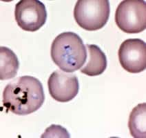
Figure A
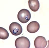
Figure B
Answer to Case #1
This was a case of babesiosis caused by Babesia microti. Diagnostic features observed were:
- pleomorphic ring-like parasites that varied in size and shape (black arrows in Figures A and B).
- dividing forms (red arrow in Figure A). Two, maybe three parasites are budding out.
- no visible pigment, even in the larger/older parasites. (Note: In Figure B, there are some dark reflections that could be mistaken for pigment; but it is a single dot at the periphery only, which rules out pigment).
If the objects were Plasmodium, they would be dividing as schizonts, with a typical morphology, and the older parasites would have visible pigment. In this patient, the diagnosis of transfusion-induced Babesia microti (based on morphology and history) was confirmed by serology and PCR performed at CDC.
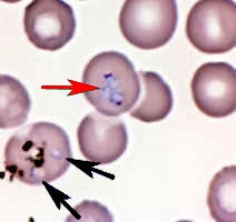
Figure A
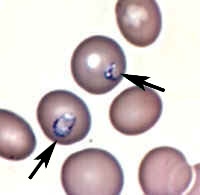
Figure B
More on: Babesiosis
Images presented in the monthly case studies are from specimens submitted for diagnosis or archiving. On rare occasions, clinical histories given may be partly fictitious.
