|
|
|
|
|
|
|
| ||||||||||
|
|
|
|
|
|
|
||||
| ||||||||||
|
|
|
|
|
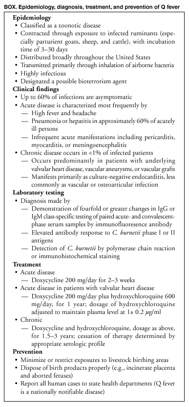
Persons using assistive technology might not be able to fully access information in this file. For assistance, please send e-mail to: mmwrq@cdc.gov. Type 508 Accommodation and the title of the report in the subject line of e-mail. Q Fever --- California, Georgia, Pennsylvania, and Tennessee, 2000--2001Q fever is a zoonotic disease caused by the bacterium Coxiella burnetii. The most common reservoirs are domesticated ruminants, primarily cattle, sheep, and goats. Humans acquire Q fever typically by inhaling aerosols or contaminated dusts derived from infected animals or animal products. Its highly infectious nature and aerosol route of transmission make C. burnetii a possible agent of bioterrorism (1). Although up to 60% of initial infections are asymptomatic (2), acute disease can manifest as a relatively mild, self-limited febrile illness, or more moderately severe disease characterized by hepatitis or pneumonia. It manifests less commonly as myocarditis, pericarditis, and meningoencephalitis. Chronic Q fever occurs in <1% of infected patients, months or years after initial infection. Chronic disease manifests most commonly as a culture-negative endocarditis in patients with valvular heart disease. During 2000--2001, a total of 48 patients who met the case definition* of Q fever were reported to CDC. This report describes the case investigations for six of these patients, which indicate that these persons acquired Q fever probably through direct or indirect contact with livestock. To enhance surveillance efforts, health-care providers should report cases of Q fever to state health departments. CaliforniaIn May 2001, a woman aged 56 years sought treatment from her health-care provider for fever (104º F [40º C]), hepatomegaly, and elevated liver enzymes (alkaline phosphatase 532 U/L [normal: 30--100 U/L], SGOT 178 U/L [normal: 9--25 U/L], and SGPT 149 U/L [normal: 7--30 U/L]). Acute cholecystitis was diagnosed, and a cholecystectomy was performed. After the procedure, the patient's symptoms persisted, and she developed pain and partial paralysis of the left leg. Approximately 4 weeks after the woman sought treatment initially, a computed tomography (CT) scan of the patient's chest revealed nonspecific interstitial lung disease. Serum samples obtained near the time of the CT scan and 6 weeks later were tested by an indirect immunofluoresence antibody (IFA) assay and demonstrated IgG antibodies reactive with C. burnetii phase II antigens at reciprocal titers of >1,024, confirming a diagnosis of Q fever. The patient's husband aged 62 years also developed a nonspecific febrile illness 3 days after the onset of his wife's illness; serum specimens obtained from him in June and July and tested by IFA demonstrated IgG antibodies reactive with C. burnetii phase II antigens at reciprocal titers of >1,024. Canvassing of the neighborhood by a public health nurse revealed that a next-door neighbor aged 76 years had a nonspecific febrile illness in April 2001. His serum was obtained in August and October and was tested by IFA; both specimens demonstrated IgG antibodies to C. burnetii phase II antigens at reciprocal titers of >1,024. The three patients were treated with doxycycline; their symptoms resolved, but the woman has residual neurologic deficits in her left leg. The couple did not own livestock but drove daily on an unpaved road past a neighbor's goat herd. Goat kids had been born at the farm during the spring. Serum specimens obtained from 48 goats in this herd were tested by CDC by using IFA; 45 (94%) animals had IgG antibodies to C. burnetii at reciprocal titers indicative of current or previous infection (titer range: 32--16,384). GeorgiaIn March 2001, a man aged 46 years sought treatment for acute onset of fever, chills, cough, and weight loss; influenza was diagnosed. The patient's symptoms persisted, and after 2 weeks he sought further treatment at an emergency department, where influenza again was diagnosed, and he was referred to an infectious disease specialist. A serum sample was tested by IFA and reacted with C. burnetii phase II antigens at a reciprocal titer of >256. The patient was administered a 5-day course of the fluoroquinolone gatifloxacin, and symptoms resolved within 2 weeks. A convalescent-phase serum sample obtained in April and tested by IFA demonstrated an IgG reciprocal antibody titer reactive with C. burnetii phase II antigens of >16,384. The patient owned several dairy cows, but there had been no recent animal births on the premises. Two beef cattle herds of approximately 35 animals each were pastured across the road from the patient's farm. Serum was drawn from 14 cattle from these herds; two animals tested by IFA reacted with phase I or II antigens of C. burnetii at reciprocal antibody titers (16--32). PennsylvaniaIn September 2000, a man aged 90 years sought treatment for fever (101.0º F [38.3º C]) and a 4-month history of malaise and weight loss after a cholecystectomy. The patient had elevated liver enzymes (alkaline phosphatase 181 U/L [normal: 45--115 U/L] and SGOT 51 U/L [normal: 1--40 U/L]). He was admitted to the hospital for diagnostic evaluation. In 1998, the patient had undergone aortic valve replacement for culture-negative endocarditis and valvular insufficiency. A serum sample drawn in November 2000 was tested by IFA and demonstrated IgG antibodies reactive with C. burnetii phase I antigens at a reciprocal titer of >524,288. Presence of C. burnetii was demonstrated in the excised aortic heart valve tissue from 1998 when tested by immunohistochemical (IHC) staining at CDC. The patient was started on long-term doxycycline therapy in October 2000. Since electing to discontinue this therapy 1 year later, the patient has had two recurrences. He was admitted to the hospital in September 2002 for fever and hypotension. The patient had owned and operated a cattle farm but had retired from farming 30 years previously. The patient's relatives raised sheep and goats nearby, but the patient denied having contact with their animals. One relative, who raised sheep, was found to have an antibody titer reactive with C. burnetii phase I antigens but had not experienced illness. TennesseeIn February 2001, a man aged 49 years was admitted to a hospital with a right lower-extremity embolism. The patient reported a 6-month history of intermittent fever, night sweats, fatigue, and arthralgias. A heart murmur had been diagnosed 4 months previously. On admission, he had a temperature of 99.2º F (37.3º C) and leukocytosis (white blood cell count of 14.3x10⁹/L [normal: 4.5--11.0x10⁹/L]). The embolism in his leg was removed surgically. An echocardiogram after hospital admission revealed a bicuspid aortic valve with moderate stenosis and severe regurgitation, and aortic valve replacement was performed. Microscopic examination of the excised valve revealed a vegetative growth, but no bacteria or fungi were detected by histopathology or routine cultures. Serum obtained 1 week after admission was tested by IFA and demonstrated IgG antibodies reactive with C. burnetii phase I antigens at a reciprocal titer of >512, and the patient was administered doxycycline and levofloxacin. CDC detected DNA of C. burnetii in the excised aortic valve by polymerase chain reaction (PCR). The embolus removed from the patient's right leg tested positive for C. burnetii by IHC staining. The patient was discharged but was readmitted 10 days later for pericardial effusion with tamponade, which resolved after surgical intervention. The patient owned one goat and a herd of approximately 100 cattle. In February 2000, the patient had been present at the stillbirth of one calf and the premature delivery and death of a second calf. Serum samples from 24 cattle in his herd were collected in July and tested for antibodies to C. burnetii by IFA; one animal had reactivity to phase I and II antigens at a reciprocal titer of 16. Reported by: M Jay-Russell, DVM, California Dept of Health Svcs; J Douglas, Calaveras County Dept of Health, San Andreas, California. C Drenzek, DVM, Georgia Div of Public Health, Dept of Human Resources. J Stone, MD, Cumberland; D Blythe, MD, Maryland Dept of Health. A Weltman, MD, Pennsylvania Dept of Health. T Jones, MD, A Craig, MD, Tennessee Dept of Health. J McQuiston, DVM, C Paddock, MD, W Nicholson, PhD, HA Thompson, PhD, Div of Viral and Rickettsial Diseases; S Zaki, MD, National Center for Infectious Diseases; J Wright, DVM, M O'Reilly, MD, D Kirschke, MD, EIS officers, CDC. Editorial Note:These cases demonstrate acute and chronic clinical characteristics of Q fever and indicate some of the risk factors for acquiring this disease (see box). The bacterium C. burnetii is distributed widely in the United States, and human cases of Q fever have been reported from almost every state (3). Human infections are associated commonly with exposure to infected animals giving birth, especially ruminants such as sheep, cattle, and goats. Cats, dogs, wildlife, and birds also are associated occasionally with human infection (3,4). Transmission to humans usually occurs by inhalation of droplets or windborne dust containing C. burnetii (2--4). The persons whose cases are described in this report acquired Q fever probably through exposure to infected livestock. Most of the six patients had occupational contact with livestock (e.g., farming); however, some of these cases demonstrate that persons need not work in a high-risk environment or have direct animal contact to become infected with C. burnetii. In humans, the clinical presentation of Q fever varies widely. Acute Q fever might be characterized by a nonspecific febrile illness, hepatitis, or pneumonia (5). Acute cholecystitis is not known to be associated with C. burnetii infection; however, the liver manifestations observed in some patients might resemble gall bladder disease. Although one person described in this report had a peripheral neuropathy after acute infection, such symptoms are uncommon (6). Chronic Q fever might manifest months to years after initial infection, most commonly as a culture-negative endocarditis (7--9). Persons with underlying heart valve defects or prosthetic valves are at increased risk for chronic Q fever endocarditis, which might occur in up to 40% of persons with valvular heart disease following acute Q fever (9). Health-care providers should be aware of the signs and symptoms of the disease and consider laboratory testing for Q fever in patients exhibiting prolonged fever, hepatitis, atypical pneumonia, or blood culture--negative endocarditis, particularly patients whose histories suggest contact with or exposure to sheep, goats, or cattle. Q fever usually is diagnosed by evaluating paired acute- and convalescent-phase serum samples. In humans, the antibody response is directed against phase I and phase II antigens of C. burnetii. Patients with acute Q fever typically produce an antibody response primarily to C. burnetii phase II antigen; chronic C. burnetii infections typically elicit a higher antibody response to phase I antigens (10). A diagnosis of Q fever also can be confirmed by examining biopsies of affected organs by using PCR or IHC. Serologic tests may be conducted at commercial laboratories, several state health laboratories, or CDC. In animals, serologic tests for anti-bodies to C. burnetii are more difficult to interpret. Presence of antibodies might indicate previous infection with the organism but cannot be used to predict human risk (3). For treatment of acute Q fever, doxycycline is the drug of choice. Initiation of therapy is warranted in patients with disease demonstrating clinical and epidemiologic features compatible with Q fever. Because antibiotic treatment is most effective during the early phase of the illness, treatment should not be withheld pending results of confirmatory laboratory antibody tests, which provide a retrospective diagnosis (2). For patients with pre-existing valvular disease, progression of acute disease to endocarditis is best prevented by combination long-term therapy with doxycycline and hydroxychloroquine. This regimen also is recommended for patients with active Q fever endocarditis (2,9). If the infection does not resolve with antibiotic therapy, the patient might require excision and replacement of the damaged heart valve; however, this will not necessarily ensure elimination of C. burnetii, and the new valve might fail if appropriate antimicrobial treatment is not initiated or is withdrawn prematurely (5). Because its highly infectious nature and aerosol route of transmission make C. burnetii a potential agent of bioterrorism, human Q fever became a nationally notifiable disease in 1999. State health departments should report cases to CDC through the National Electronic Telecommunications System for Surveillance (NETSS) via event code 10255; to facilitate case reporting, Q fever case report forms are available at http://www.cdc.gov/ncidod/dvrd/qfever/case_rep_fm.pdf. Additional information about Q fever is available at http://www.cdc.gov/ncidod/dvrd/qfever. Acknowledgments This report is based in part on data contributed by G Rishwain, MD, E Schneider, MD, Stockton, California. W Lamm, MD, Cumberland; J Roach, MD, Maryland Dept of Health. C Black, DVM, Georgia Dept of Agriculture. W Moore, R Wilson, DVM, Tennessee Dept of Health. J Childs, ScD, J Singleton, J Sumner, J Miller, Div of Viral and Rickettsial Diseases; W Shieh, MD, National Center for Infectious Diseases; J Perz, DrPH, EIS Officer, CDC. References
* Confirmed Q fever: A clinically compatible case that is laboratory confirmed with one of the following: 1) a fourfold change in antibody tier to C. burnetii antigen by immunofluoresence antibody assay or complement fixation antibody test, 2) a positive polymerase chain reaction assay, 3) culture of C. burnetii from a clinical specimen, or 4) positive immunostaining of C. burnetii in tissue. Probable Q fever: a clinically compatible case with single supportive IgG of IgM titer as defined by the testing laboratory.
Box
Disclaimer All MMWR HTML versions of articles are electronic conversions from ASCII text into HTML. This conversion may have resulted in character translation or format errors in the HTML version. Users should not rely on this HTML document, but are referred to the electronic PDF version and/or the original MMWR paper copy for the official text, figures, and tables. An original paper copy of this issue can be obtained from the Superintendent of Documents, U.S. Government Printing Office (GPO), Washington, DC 20402-9371; telephone: (202) 512-1800. Contact GPO for current prices. **Questions or messages regarding errors in formatting should be addressed to mmwrq@cdc.gov.Page converted: 10/17/2002 |
|||||||||
This page last reviewed 10/17/2002
|