This document updates and replaces CDC's previously published
"Guideline for Prevention of Nosocomial Pneumonia" (Infect Control
1982;3:327-33, Respir Care 1983;28:221-32, and Am J Infect Control
1983;11:230-44). This revised guideline is designed to reduce the
incidence
of nosocomial pneumonia and is intended for use by personnel who
are
responsible for surveillance and control of infections in
acute-care
hospitals; the information may not be applicable in long-term-care
facilities because of the unique characteristics of such settings.
This
revised guideline addresses common problems encountered by
infection-
control practitioners regarding the prevention and control of
nosocomial
pneumonia in U.S. hospitals. Sections on the prevention of
bacterial
pneumonia in mechanically ventilated and/or critically ill
patients, care
of respiratory-therapy devices, prevention of cross-contamination,
and
prevention of viral lower respiratory tract infections (e.g.,
respiratory
syncytial virus {RSV} and influenza infections) have been expanded
and
updated. New sections on Legionnaires disease and pneumonia caused
by
Aspergillus sp. have been included. Lower respiratory tract
infection
caused by Mycobacterium tuberculosis is not addressed in this
document.
Part I, "An Overview of the Prevention of Nosocomial Pneumonia,
1994,"
provides the background information for the consensus
recommendations of
the Hospital Infection Control Practices Advisory Committee
(HICPAC) in
Part II, "Recommendations for Prevention of Nosocomial Pneumonia."
Pneumonia is the second most common nosocomial infection in the
United
States and is associated with substantial morbidity and mortality.
Most
patients who have nosocomial pneumonia are infants, young children,
and
persons greater than 65 years of age; persons who have severe
underlying
disease, immunosuppression, depressed sensorium, and/or
cardiopulmonary
disease; and persons who have had thoracoabdominal surgery.
Although
patients receiving mechanically assisted ventilation do not
represent a
major proportion of patients who have nosocomial pneumonia, they
are at
highest risk for acquiring the infection. Most bacterial nosocomial
pneumonias occur by aspiration of bacteria colonizing the
oropharynx or
upper gastrointestinal tract of the patient. Because intubation and
mechanical ventilation alter first-line patient defenses, they
greatly
increase the risk for nosocomial bacterial pneumonia. Pneumonias
caused
by Legionella sp., Aspergillus sp., and influenza virus are often
caused
by inhalation of contaminated aerosols. RSV infection usually
occurs
after viral inoculation of the conjunctivae or nasal mucosa by
contaminated hands. Traditional preventive measures for nosocomial
pneumonia include decreasing aspiration by the patient, preventing
cross-contamination or colonization via hands of personnel,
appropriate
disinfection or sterilization of respiratory-therapy devices, use
of
available vaccines to protect against particular infections, and
education of hospital staff and patients. New measures being
investigated
involve reducing oropharyngeal and gastric colonization by
pathogenic
microorganisms.
Part 1. An Overview of the Prevention of Nosocomial Pneumonia, 1994
INTRODUCTION
This document updates and replaces CDC's previously published
"Guideline
for Prevention of Nosocomial Pneumonia" (Infect Control
1982;3:327-33,
Respir Care 1983; 28:221-32, and Am J Infect Control
1983;11:230-44).
This revised guideline is designed to reduce the incidence of
nosocomial
pneumonia and is intended for use by personnel who are responsible
for
surveillance and control of infections in acute-care hospitals; the
information may not be applicable in long-term-care facilities
because of
the unique characteristics of such settings.
This revised guideline addresses common problems encountered by
infection-control practitioners regarding the prevention and
control of
nosocomial pneumonia in U.S. hospitals. Sections concerning the
prevention of bacterial pneumonia in mechanically ventilated and/or
critically ill patients, care of respiratory-therapy devices,
prevention
of cross-contamination, and prevention of viral lower respiratory
tract
infections (e.g., respiratory syncytial virus {RSV} and influenza
infections) have been expanded and updated. New sections on
Legionnaires
disease and pneumonia caused by Aspergillus sp. have been included.
Lower
respiratory tract infection caused by Mycobacterium tuberculosis is
not
addressed in this document; CDC published such recommendations
previously
(1).
Part I, "An Overview of the Prevention of Nosocomial Pneumonia,
1994,"
provides the background information for the consensus
recommendations of
the Hospital Infection Control Practices Advisory Committee
(HICPAC) in
Part II, "Recommendations for Prevention of Nosocomial Pneumonia."
HICPAC
was established in 1991 to provide advice and guidance to the
Secretary
and the Assistant Secretary for Health, U.S. Department of Health
and
Human Services; the Director, CDC; and the Director, National
Center for
Infectious Diseases (NCID), CDC, regarding the practice of hospital
infection control and strategies for surveillance, prevention, and
control of nosocomial infections in U.S. hospitals. HICPAC also
advises
CDC on periodic updating of guidelines and other policy statements
regarding prevention of nosocomial infections. This guideline is
the
first of a series of CDC guidelines being revised by HICPAC and
NCID.
This guideline can be an important resource for educating
health-care
workers (HCWs) regarding prevention and control of nosocomial
respiratory
tract infections. Because education of HCWs is the cornerstone of
an
effective infection-control program, hospitals should give high
priority
to continuing infection-control educational programs for these
personnel.
BACKGROUND
Pneumonia is the second most common nosocomial infection in the
United
States and is associated with substantial morbidity and mortality.
Most
patients who have nosocomial pneumonia are infants, young children,
and
persons greater than 65 years of age; persons who have severe
underlying
disease, immunosuppression, depressed sensorium, and/or
cardiopulmonary
disease; and persons who have had thoracoabdominal surgery.
Although
patients receiving mechanically assisted ventilation do not
represent a
major proportion of patients who have nosocomial pneumonia, they
are at
highest risk for acquiring the infection.
Most bacterial nosocomial pneumonias occur by aspiration of
bacteria
colonizing the oropharynx or upper gastrointestinal tract of the
patient.
Because intubation and mechanical ventilation alter first-line
patient
defenses, they greatly increase the risk for nosocomial bacterial
pneumonia. Pneumonias caused by Legionella sp., Aspergillus sp.,
and
influenza virus are often caused by inhalation of contaminated
aerosols.
RSV infection usually occurs after viral inoculation of the
conjunctivae
or nasal mucosa by contaminated hands.
Traditional preventive measures for nosocomial pneumonia include
decreasing aspiration by the patient, preventing
cross-contamination or
colonization via hands of HCWs, appropriate disinfection or
sterilization
of respiratory-therapy devices, use of available vaccines to
protect
against particular infections, and education of hospital staff and
patients. New measures being investigated involve reducing
oropharyngeal
and gastric colonization by pathogenic microorganisms.
BACTERIAL PNEUMONIA
Etiologic Agents
The reported distribution of etiologic agents that cause
nosocomial
pneumonia differs between hospitals because of different
patient
populations and diagnostic methods employed (2-10). In general,
however, bacteria have been the most frequently isolated
pathogens
(2-6,9,11-13). During 1986-1989, aerobic bacteria comprised at
least
73%, and fungi 4%, of isolates from sputum and tracheal
aspirates
obtained from patients who had pneumonia at the University of
Michigan
Hospitals and at hospitals participating in the National
Nosocomial
Infection Surveillance (NNIS) System; only a few anaerobic
bacteria and
no viruses were reported, probably because anaerobic and viral
cultures
were not performed routinely in the reporting hospitals
(Table_1)
(3). Similarly, cultures of bronchoscopic specimens obtained
from
mechanically ventilated patients who had pneumonia have rarely
yielded
anaerobes (5-7,9,11,14,15). Only one study, which was based
primarily
on cultures of transtracheal aspirates obtained from patients
not
receiving mechanically assisted ventilation, reported a
predominance of
anaerobes (4).
Nosocomial bacterial pneumonias are frequently polymicrobial
(4,7,9,11,
12,15-19), and gram-negative bacilli are usually the
predominant
organisms (Table_1) (2-6,9,11-13). However, Staphylococcus
aureus
(especially methicillin-resistant S. aureus) (5,7,10,15,20,21)
and
other gram-positive cocci, including Streptococcus pneumoniae
(5,7),
have emerged recently as important isolates (14). In addition,
Haemophilus influenzae has been isolated from mechanically
ventilated
patients who had pneumonia that occurred within 48-96 hours
after
intubation (3-5,12,15,22). In hospitals participating in the
NNIS,
Pseudomonas aeruginosa, Enterobacter sp., Klebsiella
pneumoniae,
Escherichia coli, Serratia marcescens, and Proteus sp.
comprised 50% of
the isolates from cultures of respiratory tract specimens
obtained from
patients for whom nosocomial pneumonia was diagnosed by using
clinical
criteria; S. aureus accounted for 16%, and H. influenzae, for
6%
(Table_1) (3). Another study reported that gram-negative
bacilli
were present in 75% of quantitative cultures of
protected-specimen
brushings (PSB) obtained from patients who had acquired
nosocomial
pneumonia after receiving mechanically assisted ventilation;
40% of
these cultures were polymicrobial (5). In another published
report, 20%
of pathogens recovered from cultures of PSB, blood, pleural
fluid, or
percutaneous lung aspirate were gram-negative bacilli in pure
culture,
and 17% were polymicrobial; however, 54% of specimens did not
yield any
microorganism, probably because the patients from whom these
cultures
were obtained had been treated with antibiotics (6).
Diagnosis
Nosocomial bacterial pneumonia has been difficult to diagnose
(7,8,16,23-32). Frequently, the criteria for diagnosis have
been fever,
cough, and development of purulent sputum, in conjunction with
radiologic evidence of a new or progressive pulmonary
infiltrate, a
suggestive Gram stain, and positive cultures of sputum,
tracheal
aspirate, pleural fluid, or blood (3,4,23,25,33-36). Although
clinical
findings in conjunction with cultures of sputum or tracheal
specimens
may be sensitive for bacterial pathogens, they are highly
nonspecific,
especially in patients receiving mechanically assisted
ventilation
(8,9,12-15,18,24-26,29,31,37-42); conversely, cultures of blood
or
pleural fluid have very low sensitivity (8,18,19,43).
Because of these problems, a group of investigators recently
formulated
consensus recommendations for standardizing methods used to
diagnose
pneumonia in clinical research studies of ventilator-associated
pneumonia (44-46). These methods involve bronchoscopic
techniques such
as quantitative culture of PSB (5,7-9,13,15,27,31,38,41,47,48),
bronchoalveolar lavage (BAL) (7,12,41,47,49-54), and protected
BAL
(pBAL) (14). The reported sensitivities of such methods have
ranged,
depending on the tests or diagnostic criteria with which they
were
compared, from 70% to 100%, and the reported specificities of
these
methods have ranged from 60% to 100%. These methods are
invasive and
might cause complications such as hypoxemia, bleeding, or
arrhythmia
(8,13,42,44,52,55,56). In addition, the sensitivity of the PSB
procedure may be decreased for patients receiving antibiotic
therapy
(9,13,27). Nonbronchoscopic (NB) procedures (e.g., NB-pBAL
{12,27,57,
58} or NB-PSB {13}, which utilize blind catheterization of the
distal
airways) and quantitative culture of endotracheal aspirate
(59,60) have
been developed recently. Of these procedures, endotracheal
aspirate
culture might be the most practical. The use of these
bronchoscopic and
nonbronchoscopic diagnostic tests could help to better define
the
epidemiology of nosocomial pneumonia, especially in patients
receiving
mechanically assisted ventilation; however, additional studies
are
needed to determine each test's applicability in daily clinical
practice.
Epidemiology
Results of the NNIS indicate that pneumonias (diagnosed on the
basis of
the CDC surveillance definition of nosocomial pneumonia)
account for
approximately 15% of all hospital-associated infections and are
the
second most common type of nosocomial infection after those of
the
urinary tract (2,61). In 1984, the overall incidence of lower
respiratory tract infection was six cases per 1,000 discharged
patients
(2). The incidence per 1,000 discharged patients ranged from
4.2 cases
in nonteaching hospitals to 7.7 in university-affiliated
hospitals,
probably reflecting institutional differences in the level of
patients'
risk for acquiring nosocomial pneumonia.
Nosocomial bacterial pneumonia often has been identified as a
postoperative infection (62,63). In the Study of the Efficacy
of
Nosocomial Infection Control, which was conducted in the 1970s,
75% of
reported cases of nosocomial bacterial pneumonia occurred in
patients
who had had a surgical operation; the risk was 38 times greater
for
patients who had thoracoabdominal procedures than for those who
had
procedures involving other body sites (63). More recent
epidemiologic
studies, including NNIS studies, have identified other subsets
of
patients at high risk for acquiring nosocomial bacterial
pneumonia.
Such patients include persons greater than 70 years of age;
persons who
have endotracheal intubation and/or mechanically assisted
ventilation,
a depressed level of consciousness (particularly those with
closed-head
injury), or underlying chronic lung disease; and persons who
have
previously had an episode of a large-volume aspiration. Other
risk
factors include 24-hour ventilator-circuit changes,
hospitalization
during the fall or winter, stress-bleeding prophylaxis with
cimetidine
(either with or without antacid), administration of
antimicrobials,
presence of a nasogastric tube, severe trauma, and recent
bronchoscopy
(6,34,35,64-74).
The NNIS has stratified the incidence density of nosocomial
pneumonia
by patients' use of mechanical ventilation and type of
intensive-care
unit (ICU). From 1986 through 1990, the median rate of
ventilator-associated pneumonia cases per 1,000 ventilator-days
ranged
from 4.7 cases in pediatric ICUs to 34.4 cases in burn ICUs
(66). In
comparison, the median rate of nonventilator-associated
pneumonia cases
per 1,000 ICU-days ranged from zero cases in pediatric and
respiratory
ICUs to 3.2 cases in trauma ICUs.
Nosocomial pneumonia has been associated with high fatality
rates.
Crude mortality rates of 20%-50% and attributable mortality
rates of
30%-33% have been reported; in one study, the number of deaths
attributed to pneumonia reflected 60% of all deaths resulting
from
nosocomial infections (17,35,74-80). Patients receiving
mechanically
assisted ventilation have higher mortality rates than do
patients not
receiving ventilation support; however, other factors (e.g.,
the
patient's underlying disease{s} and organ failure) are stronger
predictors of death in patients who have pneumonia (34,74).
Analyses of pneumonia-associated morbidity have indicated that
pneumonia could prolong hospitalization by 4-9 days (79-83); in
the
United States, a conservative estimate of the direct cost of
this
prolonged hospitalization is $1.2 billion per year (83).
Nosocomial
pneumonia is a major infection-control problem because of its
reported
frequency, associated high fatality rate, and attendant costs.
IV. Pathogenesis
Bacteria can invade the lower respiratory tract by aspiration
of
oropharyngeal organisms, inhalation of aerosols containing
bacteria,
or, less frequently, by hematogenous spread from a distant body
site
(Figure_1). In addition, bacterial translocation from the
gastrointestinal tract has been hypothesized recently as a
mechanism
for infection. Of these routes, aspiration is believed to be
the most
important for both nosocomial and community-acquired pneumonia.
In radioisotope-tracer studies, 45% of healthy adults were
found to
aspirate during sleep (84). Persons who swallow abnormally
(e.g., those
who have depressed consciousness, respiratory tract
instrumentation
and/or mechanically assisted ventilation, or gastrointestinal
tract
instrumentation or diseases) or who have just undergone surgery
are
particularly likely to aspirate (6,34,35,63,85-87).
The high incidence of gram-negative bacillary pneumonia in
hospitalized
patients might result from factors that promote colonization of
the
pharynx by gram-negative bacilli and the subsequent entry of
these
organisms into the lower respiratory tract (33,88-91). Although
aerobic
gram-negative bacilli are recovered infrequently or are found
in low
numbers in pharyngeal cultures of healthy persons (88,92), the
likelihood of colonization substantially increases in comatose
patients, in patients treated with antimicrobial agents, and in
patients who have hypotension, acidosis, azotemia, alcoholism,
diabetes
mellitus, leukocytosis, leukopenia, pulmonary disease, or
nasogastric
or endotracheal tubes in place (33,91,93,94).
Oropharyngeal or tracheobronchial colonization by gram-negative
bacilli
begins with the adherence of the microorganisms to the host's
epithelial cells (90,95-97). Adherence may be affected by
multiple
factors associated with the bacteria (e.g., presence of pili,
cilia,
capsule, or production of elastase or mucinase), host cell
(e.g.,
surface proteins and polysaccharides), and environment (e.g.,
pH and
presence of mucin in respiratory secretions) (89,90,95,98-107).
Although the exact interactions between these factors have not
been
fully elucidated, studies indicate that certain substances
(e.g.,
fibronectin) can inhibit the adherence of gram-negative bacilli
to host
cells (98,100,108). Conversely, certain conditions (e.g.,
malnutrition,
severe illness, or postoperative state) can increase adherence
of
gram-negative bacteria (89,98,102,107,109).
The stomach also might be an important reservoir of organisms
that
cause nosocomial pneumonia (34,110-114). The role of the
stomach as
such a reservoir might differ depending on the patient's
underlying
conditions and on prophylactic or therapeutic interventions
(22,111,115-118). In healthy persons, few bacteria entering the
stomach
survive in the presence of hydrochloric acid at pH less than 2
(119,120). However, when gastric pH increases from the normal
levels to
greater than or equal to 4, microorganisms are able to multiply
to high
concentrations in the stomach (117,119,121-123). This can occur
in
elderly patients (121); in patients who have achlorhydria
(119), ileus,
or upper gastrointestinal disease; and in patients receiving
enteral
feeding, antacids, or histamine-2 {H-2} antagonists
(111,117,118,
123-125). Other factors (e.g., duodeno-gastric reflux and the
presence
of bile) may contribute to gastric colonization in patients who
have
impaired intestinal motility; these other factors need further
investigation (116).
Bacteria also can enter the lower respiratory tract of
hospitalized
patients through inhalation of aerosols generated primarily by
contaminated respiratory-therapy or anesthesia-breathing
equipment
(126-129). Outbreaks related to the use of respiratory-therapy
equipment have been associated with contaminated nebulizers,
which are
humidification devices that produce large amounts of aerosol
droplets
less than 4 um via ultrasound, spinning disk, or the Venturi
mechanism
(126,129,130). When the fluid in the reservoir of a nebulizer
becomes
contaminated with bacteria, the aerosol produced may contain
high
concentrations of bacteria that can be deposited deep in the
patient's
lower respiratory tract (126,130,131). Contaminated aerosol
inhalation
is particularly hazardous for intubated patients because
endotracheal
and tracheal tubes provide direct access to the lower
respiratory
tract. In contrast to nebulizers, bubble-through or wick
humidifiers
primarily increase the water-vapor (or molecular-water) content
of
inspired gases. Although heated bubble-through humidifiers
generate
aerosol droplets, they do so in quantities that may not be
clinically
important (127,132); wick humidifiers do not generate aerosols.
Bacterial pneumonia has resulted, in rare instances, from
hematogenous
spread of infection to the lung from another infection site
(e.g.,
pneumonia resulting from purulent phlebitis or right-sided
endocarditis). Another mechanism, translocation of viable
bacteria from
the lumen of the gastrointestinal tract through epithelial
mucosa to
the mesenteric lymph nodes and to the lung, has been
demonstrated in
animal models (133). Translocation is postulated to occur in
patients
with immunosuppression, cancer, or burns (133); however, data
are
insufficient to describe this mechanism in humans (134).
V. Risk Factors and Control Measures
Several large studies have examined the potential risk factors
for
nosocomially acquired bacterial pneumonia (Table_2)
(6,34,35,135,
136). Although specific risk factors have differed between
study
populations, they can be grouped into the following general
categories:
host factors (e.g., extremes of age and severe underlying
conditions, including immunosuppression); b) factors that
enhance
colonization of the oropharynx and/or stomach by microorganisms
(e.g.,
administration of antimicrobials, admission to an ICU,
underlying
chronic lung disease, or coma); c) conditions favoring
aspiration or
reflux (e.g., endotracheal intubation, insertion of nasogastric
tube,
or supine position); d) conditions requiring prolonged use of
mechanical ventilatory support with potential exposure to
contaminated
respiratory equipment and/or contact with contaminated or
colonized
hands of HCWs; and e) factors that impede adequate pulmonary
toilet
(e.g., undergoing surgical procedures that involve the head,
neck,
thorax, or upper abdomen or being immobilized as a result of
trauma or
illness) (6,33-35,62,73, 74,135).
Oropharyngeal, Tracheal, and Gastric Colonization
The association between colonization of the oropharynx
(88,137),
trachea (138), or stomach (110,111,117,123) and
predisposition to
gram-negative bacillary pneumonia prompted efforts to
prevent
infection by using either prophylactic local application of
antimicrobial agent(s) (139,140) or local bacterial
interference
(141,142). Although early studies suggested that the first
method
(i.e., use of aerosolized antimicrobials) could eradicate
common
gram-negative pathogens from the upper respiratory tract
(138),
superinfection occurred in some patients receiving this
therapy
(139-141,143,144). The second method (i.e., bacterial
interference
{with alpha-hemolytic streptococci}) has been used
successfully by
some investigators to prevent oropharyngeal colonization by
aerobic
gram-negative bacilli (141). However, the efficacy of this
method
for general usage has not been evaluated.
In many studies, the administration of antacids and H-2
blockers for
prevention of stress bleeding in critically ill,
postoperative,
and/or mechanically ventilated patients has been associated
with
gastric bacterial overgrowth (34,112,113,
118,122,123,145-147).
Sucralfate, a cytoprotective agent that has little effect on
gastric
pH and may have bactericidal properties of its own, has been
suggested as a potential substitute for antacids and H-2
blockers
(148-150). The results of clinical trials comparing the risk
for
pneumonia in patients receiving sucralfate with that in
patients
treated with antacids and/or H-2 blockers have been variable
(112,118,147,148,151-153). In most randomized trials, ICU
patients
receiving mechanically assisted ventilation who were treated
either
with only antacids or with antacids and H-2 blockers had
increased
gastric pH, high bacterial counts in the gastric fluid, and
increased risk for pneumonia in comparison with patients
treated
with sucralfate (112,118,147,148,151). In one study of a
large
number of patients, the incidence of early-onset pneumonia
(i.e.,
onset occurring less than or equal to 4 days after
intubation) did
not differ between patient groups, but late-onset pneumonia
occurred
in 5% of 76 patients treated with sucralfate, 16% of 69
treated with
antacids, and 21% of 68 treated with an H-2 blocker (147).
Conversely, a meta-analysis of data from eight earlier
studies (154)
and a later study comparing sucralfate with ranitidine (153)
did not
indicate a strong association between nosocomial pneumonia
and drugs
that increase gastric pH. Additional studies, in which
bronchoscopy
with either PSB or BAL is used to more reliably diagnose
pneumonia,
are being conducted to compare the efficacy of sucralfate
and
ranitidine.
Selective decontamination of the digestive tract (SDD) is
another
strategy designed to prevent bacterial colonization and
lower
respiratory tract infection in mechanically ventilated
patients
(155-179). SDD is aimed at preventing oropharyngeal and
gastric
colonization with aerobic gram-negative bacilli and Candida
sp.
without altering the anaerobic flora (Table_3). Various
SDD
regimens use a combination of locally administered
nonabsorbable
antibiotic agents, such as polymyxin and an aminoglycoside
(either
tobramycin, gentamicin, or, rarely, neomycin) or a quinolone
(either
norfloxacin or ciprofloxacin) coupled with either
amphotericin B or
nystatin. The local antimicrobial preparation is applied as
a paste
to the oropharynx and administered either orally or via the
nasogastric tube four times a day. In addition, in many
studies, a
systemic (intravenous) antimicrobial (e.g., cefotaxime or
trimethoprim) is administered to the patient.
Although most studies (155-158,160-167,169,170,175-177),
including
two meta-analyses (171,178), have demonstrated a decrease in
the
rates of nosocomial respiratory infections after SDD, these
studies
have been difficult to assess because they have differed in
design
and study population and many have had short follow-up
periods
(Table_3). In most of these studies, the diagnosis of
pneumonia
was based on clinical criteria; bronchoscopy with BAL or PSB
was
used in only a few studies (159,162,173,175-177,179).
Two recently published reports of large, double-blind,
placebo-
controlled trials demonstrated no benefit from SDD
(173,174). One of
these studies, which was conducted in France, noted that the
incidence of gram-negative bacillary pneumonia decreased
significantly after SDD, but this decrease was not
accompanied by a
decrease in pneumonia from all causes (173). In the other
study, no
differences were noted between patients randomly assigned to
SDD or
placebo treatment conditions; however, both patient groups
also
received simultaneous treatment with intravenous cefotaxime
(174).
Although an earlier meta-analysis indicated a trend toward
decreased
mortality in patients administered SDD (171), a more recent
and more
extensive analysis highlights the equivocal effect of SDD on
patient
mortality, as well as the high cost of using SDD to prevent
nosocomial pneumonia or death resulting from nosocomial
pneumonia
(i.e., to prevent one case of nosocomial pneumonia, six
patients
{range: five to nine patients} would have to be administered
SDD; to
prevent one death, 23 patients {range: 13-39 patients})
(178).
Furthermore, both the development of antimicrobial
resistance and
superinfection with gram-positive bacteria and other
antibiotic-
resistant nosocomial pathogens are public health concerns
(156,158,
159,161,175,180). Thus, currently available data do not
justify the
routine use of SDD for prevention of nosocomial pneumonia in
ICU
patients. SDD may be ultimately useful for specific subsets
of ICU
patients, such as patients with trauma or severe
immunosuppression
(e.g., bone-marrow-transplant recipients).
A new approach advocated to prevent oropharyngeal
colonization in
patients receiving enteral nutrition is to reduce bacterial
colonization of the stomach by acidifying the enteral feed
(181).
Although the absence of bacteria from the stomach has been
confirmed
in patients given acidified enteral feeding, the effect on
the
incidence of nosocomial pneumonia has not been evaluated
(181).
Aspiration of Oropharyngeal and Gastric Flora
Clinically important aspiration usually occurs in patients
who a)
have a depressed level of consciousness; b) have dysphagia
resulting
from neurologic or esophageal disorders; c) have an
endotracheal
(nasotracheal or orotracheal), tracheostomal, or enteral
(nasogastric or orogastric) tube in place; and/or d) are
receiving
enteral feeding (35,84,85,182-186). Placement of an enteral
tube may
increase nasopharyngeal colonization, cause reflux of
gastric
contents, or allow bacterial migration via the tube from the
stomach
to the upper airway (183,186-188). When enteral feedings are
administered, gross contamination of the enteral solution
during
preparation (189-191) and elevated gastric pH (70,192,193)
may lead
to gastric colonization with gram-negative bacilli. In
addition,
gastric reflux and aspiration might occur because of
increased
intragastric volume and pressure (70,117,183).
Although prevention of pneumonia in such patients may be
difficult,
methods that make regurgitation less likely (e.g., placing
the
patient in a semirecumbent position {i.e., by elevating the
head of
the bed} and withholding enteral feeding if the residual
volume in
the stomach is large or if bowel sounds are not heard upon
auscultation of the abdomen) may be beneficial
(185,194-197).
Conversely, equivocal results have been obtained by a)
administering
enteral nutrition intermittently in small boluses rather
than
continuously (70,193); b) using flexible, small-bore enteral
tubes
(186,198); or c) placing the enteral tube below the stomach
(e.g.,
in the jejunum) (199,200).
Mechanically Assisted Ventilation and Endotracheal
Intubation
Patients receiving continuous, mechanically assisted
ventilation
have 6-21 times the risk for acquiring nosocomial pneumonia
compared
with patients not receiving ventilatory support
(34,63,65,75). One
study indicated that the risk for developing
ventilator-associated
pneumonia increased by 1% per day (5). This increased risk
was
attributed partially to carriage of oropharyngeal organisms
upon
passage of the endotracheal tube into the trachea during
intubation,
as well as to depressed host defenses secondary to the
patient's
severe underlying illness (6,34,35,201). In addition,
bacteria can
aggregate on the surface of the tube over time and form a
glycocalyx
(i.e., a biofilm) that protects the bacteria from the action
of
antimicrobial agents or host defenses (202). Some
researchers
believe that these bacterial aggregates can become dislodged
by
ventilation flow, tube manipulation, or suctioning and
subsequently
embolize into the lower respiratory tract and cause focal
pneumonia
(203,204). Removing tracheal secretions by gentle suctioning
and
using aseptic techniques to reduce cross-contamination to
patients
from contaminated respiratory therapy equipment or
contaminated or
colonized hands of HCWs have been used traditionally to help
prevent
pneumonia in patients receiving mechanically assisted
ventilation.
The risk for pneumonia also is increased by the direct
access of
bacteria to the lower respiratory tract, which often occurs
because
of leakage around the endotracheal cuff (86,205), thus
enabling
pooled secretions above the cuff to enter the trachea (206).
In one
study, the occurrence of nosocomial pneumonia was delayed
and
decreased in intubated patients whose endotracheal tubes had
a
separate dorsal lumen that allowed drainage (i.e., by
suctioning) of
secretions in the space above the endotracheal tube cuff and
below
the glottis (206). However, additional studies are needed to
determine the cost-benefit ratio of using this device.
Cross-Colonization Via Hands of HCWs
Pathogens that cause nosocomial pneumonia (e.g.,
gram-negative
bacilli and S. aureus) are ubiquitous in hospitals,
especially in
intensive- or critical-care areas (207,208). Transmission of
these
microorganisms to patients frequently occurs via an
attending HCW's
hands that have become contaminated or transiently colonized
with
the microorganisms (209-215). Procedures such as tracheal
suctioning
and manipulation of the ventilator circuit or endotracheal
tubes
increase the opportunity for cross-contamination (215,216).
The risk
for cross-contamination can be reduced by using aseptic
techniques
and sterile or disinfected equipment when appropriate (65)
and by
eliminating pathogens from the hands of HCWs
(65,215,217-219).
In theory, adequate handwashing is an effective way of
removing
transient bacteria from the hands (218,219); however,
personnel
compliance with handwashing recommendations has been
generally poor
(220-223). For this reason, the routine use of gloves has
been
advocated to help prevent cross-contamination (224,225). The
routine
use of gloves, in addition to the use of gowns, was
associated with
a decrease in the incidence of nosocomial RSV infection
(226) and
other infections acquired in ICUs (227). However, nosocomial
pathogens can colonize gloves (228), and outbreaks have been
traced
to HCWs who did not change gloves after having contact with
one
patient and before providing care to another (229,230). In
addition,
gloved hands can be contaminated through leaks in the gloves
(231).
Contamination of Devices Used on the Respiratory Tract
Devices used on the respiratory tract for respiratory
therapy (e.g.,
nebulizers), diagnostic examination (e.g., bronchoscopes and
spirometers), and administration of anesthesia are potential
reservoirs and vehicles for infectious microorganisms
(65,232-236).
Routes of transmission might be from device to patient
(127,129,
234-244), from one patient to another (245,246), or from one
body
site to the lower respiratory tract of the same patient via
hand or
device (233,246-248). Contaminated reservoirs of
aerosol-producing
devices (e.g., nebulizers) can allow the growth of
hydrophilic
bacteria that subsequently can be aerosolized during use of
the
device (126,129,130,242). Gram-negative bacilli (e.g.,
Pseudomonas
sp., Xanthomonas sp., Flavobacterium sp., Legionella sp.,
and
nontuberculous mycobacteria) can multiply to substantial
concentrations in nebulizer fluid (241,249-251) and increase
the
risk for pneumonia in patients using such devices
(127-130,241,242,
252,253).
Proper cleaning and sterilization or disinfection of
reusable
equipment are important components of a program to reduce
infections
associated with respiratory therapy and anesthesia equipment
(234,
235,237-240,242,254-259). Many devices or parts of devices
used on
the respiratory tract have been categorized as semicritical
in the
Spaulding classification system for appropriate
sterilization or
disinfection of medical devices because they come into
direct or
indirect contact with mucous membranes but do not ordinarily
penetrate body surfaces (Appendix A), and the associated
risk for
infection in patients after the use of such devices is less
than
that associated with devices that penetrate normally sterile
tissues
(260). Thus, if sterilization of these devices by steam
autoclave or
ethylene oxide is not possible or cost-effective (261), they
can be
subjected to high-level disinfection by pasteurization at 75
C for
30 minutes (262-265) or by use of liquid chemical
disinfectants
approved by the Environmental Protection Agency (EPA) as
sterilants/disinfectants and approved for use on medical
instruments
by the Food and Drug Administration (225, 266-268).
If a respiratory device needs rinsing to remove a residual
liquid
chemical sterilant/disinfectant after chemical disinfection,
sterile
water is preferred because tap or locally prepared distilled
water
might contain microorganisms that can cause pneumonia
(249,250,
269-272). In some hospitals, a tap-water rinse followed by
air-
drying with or without an alcohol rinse (i.e., to hasten
drying) is
used (273). In theory, if complete drying is achieved after
a
tap-water rinse, the risk for nosocomial pneumonia
associated with
the use of the device is probably low. Air drying reduces
the level
of microbial contamination of the hands of HCWs after
washing, and
air drying also reduces contamination of gastrointestinal
endoscopes
(274-276). However, many semicritical items used on the
respiratory
tract (e.g., corrugated tubing, jet or ultrasonic
nebulizers, and
bronchoscopes) are difficult to dry, and the degree of
dryness of a
device is difficult to assess (265). Data are insufficient
regarding
the safety of routinely using tap water for rinsing
(followed by
drying) reusable semicritical respiratory devices after
their
disinfection or between their uses on the same patient
(242,258,273,
277).
Mechanical Ventilators, Breathing Circuits, Humidifiers,
Heat-Moisture Exchangers, and In-Line Nebulizers
Mechanical ventilators. The internal machinery of
mechanical
ventilators used for respiratory therapy is not
considered an
important source of bacterial contamination of inhaled
gas
(278). Thus, routine sterilization or high-level
disinfection
of the internal machinery is considered unnecessary.
Using
high-efficiency bacterial filters at various positions
in the
ventilator breathing circuit had been advocated
previously
(279,280). Filters interposed between the machinery
and the
main breathing circuit can eliminate contaminants from
the
driving gas and prevent retrograde contamination of
the
machine by the patient; however, these filters also
might
alter the functional specifications of the breathing
device
by impeding high gas flows (279-281). Placement of a
filter
or condensate trap at the expiratory-phase tubing of
the
mechanical-ventilator circuit may help prevent cross-
contamination of the ventilated patient's immediate
environment (247,282), but the importance of such
filters in
preventing nosocomial pneumonia needs further
evaluation.
Breathing circuits, humidifiers, and heat-moisture
exchangers. In the United States, most hospitals use
ventilators with either bubble-through or wick
humidifiers
that produce either insignificant (132,283) or no
aerosols,
respectively, for humidification. Thus, these devices
probably do not pose an important risk for pneumonia
in
patients. In addition, bubble-through humidifiers are
usually
heated to temperatures that reduce or eliminate
bacterial
pathogens (283,284). Sterile water, however, is still
usually
used to fill these humidifiers (285) because tap or
distilled
water might contain microorganisms, such as Legionella
sp.,
that are more heat-resistant than other bacteria
(252,271).
The potential risk for pneumonia in patients using
mechanical
ventilators that have heated bubble-through
humidifiers stems
primarily from the condensate that forms in the
inspiratory-
phase tubing of the ventilator circuit as a result of
the
difference in the temperatures of the
inspiratory-phase gas
and ambient air; condensate formation increases if the
tubing
is unheated (286). The tubing and condensate can
rapidly
become contaminated, usually with bacteria that
originate in
the patient's oropharynx (286). In one study, 33% of
inspiratory circuits were colonized with bacteria via
this
route within 2 hours, and 80% within 24 hours, after
initiation of mechanical ventilation (286). Spillage
of the
contaminated condensate into the patient's
tracheobronchial
tree, as can occur during procedures in which the
tubing is
moved (e.g., for suctioning, adjusting the ventilator
setting, or feeding or caring for the patient), may
increase
the risk for pneumonia in the patient (286). Thus, in
many
hospitals, HCWs are trained to prevent such spillage
and to
drain the fluid periodically. Microorganisms
contaminating
ventilator-circuit condensate can be transmitted to
other
patients via the hands of HCWs handling the fluid,
especially
if the HCW neglects washing hands after handling the
condensate.
The role of ventilator-tubing changes in preventing
pneumonia
in patients using mechanical ventilators with
bubble-through
humidifiers has been investigated. Initial studies of
in-use
contamination of mechanical ventilator circuits with
humidifiers have indicated that neither the rate of
bacterial
contamination of inspiratory-phase gas nor the
incidence of
pneumonia was significantly increased when tubing was
changed
every 24 hours rather than every 8 or 16 hours (287).
A later
study indicated that changing the ventilator circuit
every 48
hours rather than every 24 hours did not result in an
increase in contamination of the inspiratory-phase gas
or
tubing of the ventilator circuits (288). In addition,
the
incidence of nosocomial pneumonia was not
significantly
higher when circuits were changed every 48 hours
rather than
every 24 hours (288). More recent reports suggest that
the
risk for pneumonia may not increase when the interval
for
circuit change is prolonged beyond 48 hours. Another
study
indicated that the risk for pneumonia was not
significantly
higher when the circuits were never changed for the
duration
of use by the patient (eight {29%} of 28 patients)
rather
than when the circuits were changed every 48 hours (11
{31%}
of 35 patients) (289).
These findings indicate that the recommended daily
change in
ventilator circuits may be extended to greater than or
equal
to 48 hours. This change in recommendation could
result in
substantial savings for U.S. hospitals by reducing the
number
of circuits used and the amount of personnel time
required to
change the circuits (285,288). The maximum time,
however,
that a circuit can be safely left unchanged on a
patient has
not been determined.
Condensate formation in the inspiratory-phase tubing
of a
ventilator breathing circuit can be decreased by
elevating
the temperature of the inspiratory-phase gas with a
heated
wire in the inspiratory-phase tubing. However, in one
report,
three cases of endotracheal- or tracheostomy-tube
blockage by
dried secretions of the patient were attributed to the
decrease in the relative humidity of inspired gas that
resulted from the elevation of the gas temperature
(290).
Until additional information regarding the frequency
of such
cases is available, HCWs who provide care to patients
requiring mechanical ventilation should be aware of
the
advantages and potential complications associated with
using
heated ventilator tubing.
Condensate formation can be eliminated by using a
heat-moisture exchanger (HME) or a hygroscopic
condenser
humidifier (i.e., an "artificial nose") (291-296). An
HME
recycles heat and moisture exhaled by the patient and
eliminates the need for a humidifier. In the absence
of a
humidifier, no condensate forms in the
inspiratory-phase
tubing of the ventilator circuit. Thus, bacterial
colonization of the tubing is prevented, and the need
to
change the tubing on a periodic basis is obviated
(216). Some
models of HMEs are equipped with bacterial filters,
but the
advantage of using such filters is unknown. HMEs can
increase
the dead space (i.e., the area of the lung in which
air is
not exchanged) and resistance to breathing, might leak
around
the endotracheal tube, and might result in drying of
sputum
and blockage of the tracheobronchial tree (297).
Although
recently developed HMEs that have humidifiers increase
airway
humidity without increasing colonization of bacteria
(293,
298), additional studies are needed to determine
whether the
incidence of pneumonia is decreased (299-302).
Small-volume ("in-line") medication nebulizers.
Small-volume
medication nebulizers that are inserted in the
inspiratory
circuit of mechanical ventilators can produce
bacterial
aerosols (242). If such devices become contaminated by
condensate in the inspiratory tubing of the breathing
circuit, they can increase the patient's risk for
pneumonia
because the nebulizer aerosol is directed through the
endotracheal tube and bypasses many of the normal host
defenses against infection (286).
Large-Volume Nebulizers. Nebulizers with large-volume
(greater
than 500 cc) reservoirs, including those used in
intermittent
positive-pressure breathing (IPPB) machines and
ultrasonic or
spinning-disk room-air humidifiers, pose the greatest
risk for
pneumonia to patients, probably because of the large
amount of
aerosols they generate (237-241,252,303). These
reservoirs can
become contaminated by the hands of HCWs, unsterile
humidification fluid, or inadequate sterilization or
disinfection
between uses (126). Once introduced into the reservoir,
various
bacteria, including Legionella sp., can multiply to
sufficiently
large numbers within 24 hours to pose a risk for
infection in
patients who receive inhalation therapy
(128,129,241,253,303).
Sterilization or high-level disinfection of these
nebulizers can
eliminate vegetative bacteria from their reservoirs and
make them
safe for patient use (260). However, unlike nebulizers
attached
to IPPB machines, room-air humidifiers have a high
cost-benefit
ratio: evidence of clinical benefits from their use in
hospitals
is lacking, and the potential cost of daily sterilization
or
disinfection of, and use of sterile water to fill, such
devices
is substantial.
Hand-Held Small-Volume Medication Nebulizers.
Small-volume
medication nebulizers used to administer bronchodilators,
including nebulizers that are hand-held, can produce
bacterial
aerosols. Hand-held nebulizers have been associated with
nosocomial pneumonia, including Legionnaires disease,
resulting
from either contamination with medications from multidose
vials
(304) or Legionella-contaminated tap water used for
rinsing and
filling the reservoir (258).
Suction Catheters, Resuscitation Bags, Oxygen Analyzers,
and
Ventilator Spirometers. Tracheal suction catheters can
introduce
microorganisms into a patient's lower respiratory tract.
Two
types of suction-catheter systems are used in U.S.
hospitals: the
open single-use catheter system and the closed multi-use
catheter
system. Studies comparing the two systems have involved
low
numbers of patients; the results of these studies suggest
that
the risk for catheter contamination or pneumonia does not
differ
between patients on whom the single-use suction method is
used
and those on whom the closed multi-use catheter system is
used
(305-307). Although advantages of cost and decreased
environmental contamination have been attributed to use
of the
closed-suction system (308,309), larger studies are
needed to
compare the advantages and disadvantages of both systems
(310).
Reusable resuscitation bags are particularly difficult to
clean
and dry between uses; microorganisms in secretions or
fluid left
in the bag may be aerosolized and/or sprayed into the
lower
respiratory tract of the patient on whom the bag is used;
in
addition, contaminating microorganisms might be
transmitted from
one patient to another via hands of HCWs (311-313).
Oxygen
analyzers and ventilator spirometers have been associated
with
outbreaks of gram-negative respiratory tract colonization
and
pneumonia resulting from patient-to-patient transmission
of
organisms via hands of HCWs (233,245). These devices
require
either sterilization or high-level disinfection between
uses on
different patients. Education of physicians, respiratory
therapists, and nursing staff regarding the associated
risks and
appropriate care of these devices is essential.
Anesthesia Equipment. The contributory role of anesthesia
equipment in outbreaks of nosocomial pneumonia was
reported
before hospitals implemented routine after-use cleaning
and
disinfection/sterilization of reusable
anesthesia-equipment
components that could become contaminated with pathogens
during
use (314,315).
Anesthesia machine. The internal components of
anesthesia
machines, which include the gas sources and outlets,
gas
valves, pressure regulators, flowmeters, and
vaporizers, are
not considered an important source of bacterial
contamination
of inhaled gases (316). Thus, routine sterilization or
high-level disinfection of the internal machinery is
unnecessary.
Breathing system or patient circuit. The breathing
system or
patient circuit (including the tracheal tube or face
mask,
inspiratory and expiratory tubing, y-piece, CO2
absorber and
its chamber, anesthesia ventilator bellows and tubing,
humidifier, adjustable pressure-limiting valve, and
other
devices and accessories), through which inhaled and/or
exhaled gases flow to and from a patient, can become
contaminated with microorganisms that might originate
from
the patient's oropharynx or trachea. Recommendations
for
in-use care, maintenance, and reprocessing (i.e.,
cleaning
and disinfection or sterilization) of the components
of the
breathing system have been published (317,318). In
general,
reusable components of the breathing system that
directly
touch the patient's mucous membranes (e.g., face mask
or
tracheal tube) or become readily contaminated with the
patient's respiratory secretions (e.g., y-piece,
inspiratory
and expiratory tubing, and attached sensors) are
cleaned and
subjected to high-level disinfection or sterilization
between
patients. The other parts of the breathing system
(e.g., CO2
absorber and its chamber), for which an appropriate
and
cost-effective schedule of reprocessing has not been
firmly
determined (319), are changed, cleaned, and sterilized
or
subjected to high-level disinfection periodically in
accordance with published guidelines (317,318) and/or
the
manufacturers' instructions.
Using high-efficiency bacterial filters at various
positions
in the patient circuit (e.g., at the y-piece or on the
inspiratory and expiratory sides of the patient
circuit) has
been advocated (317,320,321) and shown to decrease
contamination of the circuit (321-323). However, the
use of
bacterial filters to prevent nosocomial pulmonary
infections
has not been proven to be effective and requires
additional
analysis (324-326).
Pulmonary Function Testing Apparatus.
Internal parts of pulmonary function testing
apparatus. The
internal parts of pulmonary function testing apparatus
usually are not considered an important source of
bacterial
contamination of inhaled gas (327). However, because
of
concern about possible carry-over of bacterial
aerosols from
an infectious patient-user of the apparatus to the
next
patient (246,328), placement of bacterial filters
(i.e., that
remove exhaled bacteria) between the patient and the
testing
equipment has been advocated (246,329). More studies
are
needed to evaluate the need for and efficacy of these
filters
in preventing nosocomial pneumonia (330).
Tubing, rebreathing valves, and mouthpieces. Tubing,
connectors, rebreathing valves, and mouthpieces could
become
contaminated with patient secretions during use of the
pulmonary function testing apparatus. Thus, these
items
should be cleaned and subjected to high-level
disinfection or
sterilization between uses on different patients.
Thoracoabdominal Surgical Procedures
Certain patients are at high risk for developing
postoperative
pulmonary complications, including pneumonia. These persons
include
those who are obese or are greater than 70 years of age or
who have
chronic obstructive pulmonary disease (331-334). Abnormal
results
from pulmonary function tests (especially decreased maximum
expiration flow rate), a history of smoking, the presence of
tracheostomy or prolonged intubation, or protein depletion
that can
cause respiratory-muscle weakness are also risk factors
(62,68,136).
Patients who undergo surgery of the head, neck, thorax, or
abdomen
might have impairment of normal swallowing and respiratory
clearance
mechanisms as a result of instrumentation of the respiratory
tract,
anesthesia, or increased use of narcotics and sedatives
(332,335,
336). Patients who undergo upper abdominal surgery usually
have
diaphragmatic dysfunction that results in decreased
functional
residual capacity of the lungs, closure of airways, and
atelectasis
(337,338).
Interventions aimed at reducing the postoperative patient's
risk for
pneumonia have been developed (339). These include deep
breathing
exercises, chest physiotherapy, use of incentive spirometry,
IPPB,
and continuous positive airway pressure by face mask
(339-349).
Studies evaluating the relative efficacy of these modalities
reported variable results and were difficult to compare
because of
differences in outcome variables assessed, patient
populations
studied, and study design (339,341,342,348-350).
Nevertheless, many
studies have reported that deep breathing exercises, use of
incentive spirometry, and IPPB are advantageous maneuvers,
especially in patients who had preoperative pulmonary
dysfunction
(342,343,345,346,348-350). In addition, control of pain that
interferes with cough and deep breathing during the
immediate
postoperative period decreases the incidence of pulmonary
complications after surgery. Several methods of controlling
pain
have been used; these include both intramuscular or
intravenous
(including patient-controlled) administration of analgesia
and
regional (e.g., epidural) analgesia (351-358).
Other Prophylactic Measures
Vaccination of Patients. Although pneumococci are not a
major
cause of nosocomial pneumonia, these organisms have been
identified as etiologic agents of serious nosocomial
pulmonary
infection and bacteremia (359-361). The following factors
place
patients at high risk for complications from pneumococcal
infections: age greater than or equal to 65 years of age,
chronic
cardiovascular or pulmonary disease, diabetes mellitus,
alcoholism, cirrhosis, cerebrospinal fluid leaks,
immunosuppression, functional or anatomic asplenia, or
infection
with human immunodeficiency virus (HIV). Pneumococcal
vaccine is
effective in preventing pneumococcal disease (362,363).
Because
two thirds or more of patients with serious pneumococcal
disease
have been hospitalized at least once within the 5 years
preceding
their pneumococcal illness, offering pneumococcal vaccine
in
hospitals (e.g., at the time of patient discharge) should
contribute substantially to preventing the disease
(362,364).
Prophylaxis with Systemic Antimicrobial Agents. The
systemic
administration of antimicrobials is commonly used to
prevent
nosocomial pneumonia -- especially for patients who are
receiving
mechanical ventilation, are postoperative, and/or are
critically
ill (365-367). However, the efficacy of this practice is
questionable, and superinfection, which is possible as a
result
of any antimicrobial therapy, could occur
(74,91,366-371).
Use of "Kinetic Beds" or Continuous Lateral Rotational
Therapy
(CLRT) for Immobilized Patients. Use of kinetic beds, or
CLRT, is
a maneuver for prevention of pulmonary and other
complications
resulting from prolonged immobilization or bed rest, such
as in
patients with acute stroke, critical illness, head injury
or
traction, blunt chest trauma, and/or mechanically
assisted
ventilation (372-377). This procedure involves the use of
a bed
that turns continuously and slowly (from less than or
equal to 40
for CLRT to greater than or equal to 40 for kinetic
therapy)
along its longitudinal axis. Among the hypothesized
benefits are
improved drainage of secretions within the lungs and
lower
airways, increased tidal volume, and reduction of venous
thrombosis with resultant pulmonary embolization
(378-381).
However, the efficacy of CLRT in preventing pneumonia
needs
further evaluation because studies have yielded variable
results
(372-376). In addition, the studies either involved small
numbers
of patients (373), lacked adequate randomization (372),
had no
clear definition of pneumonia (372), did not distinguish
between
community-acquired and nosocomial pneumonia (373,377), or
did not
adjust for possible confounding factors (e.g., mechanical
ventilation, endotracheal intubation, nasogastric
intubation, and
enteral feeding) (372).
LEGIONNAIRES DISEASE
Epidemiology
Legionnaires disease is a multisystem illness, with pneumonia,
caused
by Legionella sp. (382). Since the etiologic agent of
Legionnaires
disease was identified, numerous nosocomial outbreaks of the
disease
have been reported, thus enabling researchers to study the
epidemiology
of epidemic legionellosis. In contrast, the epidemiology of
sporadic
(i.e., nonoutbreak-related) nosocomial Legionnaires disease has
not
been well defined. However, when one case is identified, the
presence
of additional cases should be suspected. Of 196 cases of
nosocomial
Legionnaires disease reported in England and Wales during
1980-1992,
69% occurred during 22 nosocomial outbreaks (defined as two or
more
cases occurring at a hospital during a 6-month period) (383).
Nine
percent of cases occurred greater than 6 months before or after
a
hospital outbreak, and another 13% occurred in hospitals in
which other
sporadic cases, but no outbreaks, were identified. Only 9%
occurred at
institutions in which no outbreaks or additional sporadic cases
were
identified.
In North America, the overall proportion of nosocomial
pneumonias
caused by Legionella sp. has not been determined, although the
reported
proportions from individual hospitals have ranged from zero to
14%
(384-386). Because diagnostic tests for Legionella sp.
infection are
not performed routinely on all patients who have
hospital-acquired
pneumonia in most U.S. hospitals, this range probably
underestimates
the incidence of Legionnaires disease.
Legionella sp. are commonly found in various natural and
man-made
aquatic environments (387,388) and may enter hospital water
systems in
low or undetectable numbers (389,390). Cooling towers,
evaporative
condensers, heated potable-water-distribution systems within
hospitals,
and locally produced distilled water can provide a suitable
environment
for legionellae to multiply. Factors known to enhance
colonization and
amplification of legionellae in man-made water environments
include
temperatures of 25-42 C (391-395), stagnation (396), scale and
sediment
(392), and the presence of certain free-living aquatic amoebae
that are
capable of supporting intracellular growth of legionellae
(397,398).
A person's risk for acquiring legionellosis after exposure to
contaminated water depends on a number of factors, including
the type
and intensity of exposure and the person's health status
(399-401).
Persons who are severely immunosuppressed or who have chronic
underlying illnesses, such as hematologic malignancy or
end-stage renal
disease, are at a markedly increased risk for legionellosis
(401-404).
Persons in the later stages of acquired immunodeficiency
syndrome
(AIDS) also are probably at increased risk for legionellosis,
but data
are limited because of infrequent testing of patients (401).
Persons
who have diabetes mellitus, chronic lung disease, or
nonhematologic
malignancy; those who smoke cigarettes; and the elderly are at
moderately increased risk (382). Nosocomial Legionnaires
disease also
has been reported among patients in pediatric hospitals
(405,406).
Underlying disease and advanced age are risk factors not only
for
acquiring Legionnaires disease but also for dying as a result
of the
illness. In a multivariate analysis of 3,524 cases reported to
CDC from
1980 through 1989, immunosuppression, advanced age, end-stage
renal
disease, cancer, and nosocomial acquisition of disease were
each
independently associated with a fatal outcome (401). The
mortality rate
was 40% among 803 persons who had nosocomially acquired cases,
compared
with 20% among 2,721 persons who had community-acquired cases
(401);
this difference probably reflected the increased severity of
underlying
disease in hospitalized patients.
Diagnosis
The clinical spectrum of disease caused by Legionella sp. is
broad and
ranges from asymptomatic infection to rapidly progressive
pneumonia.
Legionnaires disease cannot be distinguished clinically or
radiographically from pneumonia caused by other agents
(407,408), and
evidence of infection with other respiratory pathogens does not
exclude
the possibility of concomitant Legionella sp. infection
(409-411).
The diagnosis of legionellosis may be confirmed by any one of
the
following: culture isolation of Legionella from respiratory
secretions
or tissues, microscopic visualization of the bacterium in
respiratory
secretions or tissue by immunofluorescent microscopy, or, for
legionellosis caused by Legionella pneumophila serogroup 1,
detection
of L. pneumophila serogroup-1 antigens in urine by
radioimmunoassay, or
observation of a four-fold rise in L. pneumophila serogroup-1
antibody
titer to greater than or equal to 1:128 in paired acute and
convalescent serum specimens by use of an indirect
immunofluorescent
antibody (IFA) test (412,413). A single elevated antibody titer
does
not confirm a case of Legionnaires disease because IFA titers
greater
than or equal to 1:256 are found in 1%-16% of healthy adults
(410,
414-417).
Because the above tests complement each other, performing each
test
when Legionnaires disease is suspected increases the
probability of
confirming the diagnosis (418). However, because none of the
laboratory
tests is 100% sensitive, the diagnosis of legionellosis is not
excluded
even if one or more of the tests are negative (413,418). Of the
available tests, the most specific is culture isolation of
Legionella
sp. from any respiratory tract specimen (419,420).
Modes of Transmission
Inhalation of aerosols of water contaminated with Legionella
sp. might
be the primary mechanism by which these organisms enter a
patient's
respiratory tract (382). In several hospital outbreaks,
patients were
considered to be infected through exposure to contaminated
aerosols
generated by cooling towers, showers, faucets, respiratory
therapy
equipment, and room-air humidifiers (11,241,258, 421-427). In
other
studies, aspiration of contaminated potable water or pharyngeal
colonizers was proposed as the mode of transmission to certain
patients
(425,428-430). However, person-to-person transmission has not
been
observed.
IV. Definition of Nosocomial Legionnaires Disease
The incubation period for Legionnaires disease is usually 2-10
days
(431); thus, for the purposes of this document and the
accompanying
HICPAC recommendations, laboratory-confirmed legionellosis that
occurs
in a patient who has been hospitalized continuously for greater
than or
equal to 10 days before the onset of illness is considered a
definite
case of nosocomial Legionnaires disease, and
laboratory-confirmed
infection that occurs 2-9 days after hospital admission is a
possible
case of the disease.
V. Prevention and Control Measures
Prevention of Legionnaires Disease in Hospitals with No
Identified
Cases (Primary Prevention)
Prevention strategies in health-care facilities in which no
cases of
nosocomial legionellosis have been identified have differed
depending on the immunologic status of the patients, the
design and
construction of the facility, the resources available for
implementing prevention strategies, and state and local
regulations.
At least two strategies are practiced with regard to the
most
appropriate and cost-effective means of preventing
nosocomial
legionellosis, especially in hospitals in which no cases or
only
sporadic cases of the illness have been detected. However, a
study
comparing the cost-benefit ratios of these strategies has
not been
conducted.
The first approach is based on periodic, routine culturing
of water
samples from the hospital's potable water system for the
purpose of
detecting Legionella sp. (432,433). When greater than or
equal to
30% of the samples obtained are culture-positive for
Legionella sp.,
the hospital's potable water system is decontaminated (433),
and
diagnostic laboratory tests for legionellosis are made
available to
clinicians in the hospital's microbiology department so that
active
surveillance for cases can be implemented (433,434). This
approach
is based on the premise that no cases of nosocomial
legionellosis
can occur if Legionella sp. is not present in the potable
water
system, and, conversely, if Legionella sp. are cultured from
the
water, cases of nosocomial legionellosis could occur
(428,435).
Proponents of this strategy indicate that when physicians
are
informed that the potable water system of the hospital is
culture-positive for Legionella sp., they are more inclined
to
conduct the necessary tests for legionellosis (434). A
potential
advantage of using this approach in hospitals in which no
cases of
nosocomial legionellosis have occurred is that routinely
culturing a
limited number of water samples is less costly than
routinely
performing laboratory diagnostic testing for all patients
who have
nosocomial pneumonia.
The main argument against this approach is that, in the
absence of
cases, the relationship between the results of water
cultures and
the risk for legionellosis remains undefined. The bacterium
has been
frequently present in water systems of buildings (436),
often
without being associated with known cases of disease
(271,385,437,
438). In a study of 84 hospitals in Quebec, 68% of the water
systems
were found to be colonized with Legionella sp., and 26% were
colonized at greater than 30% of sites sampled; however,
cases of
Legionnaires disease were reported rarely from these
hospitals
(271). Similarly, at one hospital in which active
surveillance for
legionellosis and environmental culturing for Legionella sp.
were
done, no cases of legionellosis occurred in a urology ward
during a
3.5-month period when 70% of water samples from the ward
were
culture-positive for L. pneumophila serogroup 1 (385).
Interpretation of the results of routinely culturing the
water might
be confounded by differing results among the sites sampled
within a
single water system and by fluctuations in the concentration
of
Legionella sp. at the same site (439,440). In addition, the
risk for
illness after exposure to a given source might be influenced
by a
number of factors other than the presence or concentration
of
organisms; these factors include the degree to which
contaminated
water is aerosolized into respirable droplets, the proximity
of the
infectious aerosol to the potential host, the susceptibility
of the
host, and the virulence properties of the contaminating
strain
(441-443). Thus, data are insufficient to assign a level of
risk for
disease even on the basis of the number of colony-forming
units
detected in samples from the hospital environment. By
routinely
culturing water samples, many hospital administrators will
have to
initiate water-decontamination programs if Legionella sp.
are
identified. Because of this problem, routine monitoring of
water
from the hospital's potable water system and from
aerosol-producing
devices is not widely recommended (444).
The second approach to preventing and controlling nosocomial
legionellosis involves a) maintaining a high index of
suspicion for
legionellosis and appropriately using diagnostic tests for
legionellosis in patients who have nosocomial pneumonia and
who are
at high risk for developing the disease and dying from the
infection
(385,445), b) initiating an investigation for a hospital
source of
Legionella sp. upon identification of one case of definite
or two
cases of possible nosocomial Legionnaires disease, and c)
routinely
maintaining cooling towers and using only sterile water for
the
filling and terminal rinsing of nebulization devices.
Measures used in hospitals in which cases of nosocomial
legionellosis have been identified include either a) routine
maintenance of potable water at greater than or equal to 50
C or
less than 20 C at the tap or b) chlorination of heated water
to
achieve 1-2 mg/L of free residual chlorine at the tap,
especially in
areas where immunosuppressed and other high-risk patients
are
located (385,428,439,446-449). However, the cost-benefit
ratio of
such measures in hospitals in which no cases of
legionellosis have
been identified needs additional evaluation.
Prevention of Legionnaires Disease in Hospitals with
Identified
Cases (Secondary Prevention)
The indications for a full-scale environmental investigation
to
search for and subsequently decontaminate identified sources
of
Legionella sp. in hospital environments have not been
clarified, and
these indications probably differ depending on the hospital.
In
hospitals in which as few as one to three nosocomial cases
are
identified during a period of several months, intensified
surveillance for Legionnaires disease has frequently
identified
numerous additional cases (403,422,425,447). This finding
suggests
the need for a low threshold for initiating an investigation
after
laboratory confirmation of cases of nosocomial
legionellosis.
However, when developing a strategy for responding to such
an
identification, infection-control personnel should consider
the
level of risk for nosocomial acquisition of, and mortality
from,
Legionella sp. infection at their particular hospital.
An epidemiologic investigation conducted to determine the
source of
Legionella sp. involves several important steps. First,
microbiologic and medical records should be reviewed.
Second, active
surveillance should be initiated to identify all recent or
ongoing
cases of legionellosis. Third, potential risk factors for
infection
(including environmental exposures such as showering or use
of
respiratory-therapy equipment) should be identified by
creating a
line listing of cases, analyzing the collected information
(by time,
place, and person), and comparing case-patients with
appropriate
controls. Fourth, water samples should be collected from
environmental sources implicated by the epidemiologic
investigation
and from other potential sources of aerosolized water.
Fifth,
subtype-matching between legionellae isolated from patients
and
environmental samples should be conducted (427,450-452).
This last
step can be crucial in supporting epidemiologic evidence of
a link
between human illness and a specific source (453).
In some hospitals in which the heated-water system was
identified as
the source of the organism, the system was decontaminated by
pulse
(one-time) thermal disinfection or superheating (i.e.,
flushing each
distal outlet of the hot-water system for at least 5 minutes
with
water at greater than or equal to 65 C) and
hyperchlorination
(flushing all outlets of the hot-water system with water
containing
greater than or equal to 10 mg/L of free residual chlorine)
(449,
454-456). After either of these procedures, most hospitals
either a)
maintain heated water at greater than or equal to 50 C or
less than
20 C at the tap or b) chlorinate heated water to achieve 1-2
mg/L of
free residual chlorine at the tap (385,428,439,446-449).
Additional
measures (e.g., physical cleaning or replacement of
hot-water
storage tanks, water-heaters, faucets, and showerheads) may
be
required because scale and sediment might accumulate in this
equipment and protect organisms from the biocidal effects of
heat
and chlorine (392,449). Alternative methods for controlling
and
eradicating legionellae in water systems (e.g., treating
water with
ozone, ultraviolet light, or heavy metal ions) have limited
the
growth of legionellae under laboratory and/or operating
conditions
(457-462). However, additional data are needed regarding the
efficacy of these methods before they can be considered
standard
precautions. Measures for decontaminating hospital cooling
towers
have been published previously (463).
Additional preventive measures have been used to protect
severely
immunocompromised patients. At one hospital,
immunosuppressed
patients were restricted from taking showers, and, for these
patients, only sterile water was used for drinking or
flushing
nasogastric tubes (429). In another hospital, a combined
approach
consisting of continuous heating, particulate filtration,
ultraviolet treatment, and monthly pulse hyperchlorination
of the
water supply to the bone-marrow transplant unit was used to
decrease
the incidence of Legionnaires disease (458).
The decision to search for hospital environmental sources of
Legionella sp. and the choice of procedures to use to
eradicate such
contamination should take into account the type of patient
population served by the hospital. Furthermore, decision
makers
should consider a) the high cost of an environmental
investigation
and of instituting control measures to eradicate Legionella
sp. from
sources in the hospital (464,465) and b) the differential
risk,
based on host factors, for acquiring nosocomial
legionellosis and of
having severe and fatal infection with the microorganism.
ASPERGILLOSIS
Epidemiology
Aspergillus sp. are ubiquitous fungi that commonly occur in
soil,
water, and decaying vegetation. Aspergillus sp. have been
cultured from
unfiltered air, ventilation systems, contaminated dust
dislodged during
hospital renovation and construction, horizontal surfaces,
food, and
ornamental plants (466).
Aspergillus fumigatus and Aspergillus flavus are the most
frequently
isolated Aspergillus sp. in patients who have
laboratory-confirmed
aspergillosis (467). Nosocomial aspergillosis has been
recognized
increasingly as a cause of severe illness and mortality in
highly
immunocompromised patients (e.g., patients undergoing
chemotherapy
and/or organ transplantation, including bone-marrow
transplantation for
hematologic and other malignant neoplasms) (468-472).
The most important nosocomial infection caused by Aspergillus
sp. is
pneumonia (473,474). Hospital outbreaks of pulmonary
aspergillosis have
occurred primarily in granulocytopenic patients, especially
those in
bone-marrow transplant units (473-480). Although invasive
aspergillosis
has been reported in recipients of solid-organ (e.g., heart and
kidney)
transplants (481-485), the incidence of Aspergillus sp.
infections in
these patients has been lower than in recipients of bone-marrow
transplants, probably because granulocytopenia is less severe
in
solid-organ transplant recipients and the use of
corticosteroids,
especially in kidney transplant recipients, has decreased with
the
introduction of cyclosporine (483,486). The efficacy of
infection-
control measures, such as provision of protected environments
and
prophylaxis with antifungal agents, in preventing aspergillosis
in
solid-organ transplant recipients has not been well evaluated
(483,484,486,487). In one study of heart-transplant recipients,
using
only protective isolation of patients did not prevent fungal
infections
(488).
The reported attributable mortality from invasive pulmonary
aspergillosis has differed depending on the patient population
studied.
Rates have been as high as 95% in recipients of allogeneic
bone-marrow
transplants and patients who have aplastic anemia, compared
with rates
of 13%-80% in leukemic patients (489-491).
Pathogenesis
In contrast to most bacterial pneumonias, the primary route of
acquiring Aspergillus sp. infection is by inhalation of the
fungal
spores. In severely immunocompromised patients, primary
Aspergillus sp.
pneumonia results from invasion of local lung tissue
(467,474,492).
Subsequently, the fungus might disseminate via the bloodstream
to
involve multiple other deep organs (467,474,493). A role for
nasopharyngeal colonization with Aspergillus sp., as an
intermediate
step before invasive pulmonary disease, has been proposed but
remains
to be elucidated (494-496). Conversely, colonization of the
lower
respiratory tract by Aspergillus sp. has predisposed patients,
especially those with preexisting lung disease (e.g., chronic
obstructive lung disease, cystic fibrosis, or inactive
tuberculosis),
to invasive pulmonary and/or disseminated infection
(467,474,497).
Diagnosis
Diagnosing pneumonia caused by Aspergillus sp. is often
difficult
without performing invasive procedures. Although
bronchoalveolar lavage
has been a useful screening test (498-500), lung biopsy is
still
considered the most reliable technique (501). Histopathologic
demonstration of tissue invasion by fungal hyphae has been
required in
addition to isolation of Aspergillus sp. from respiratory tract
secretions because the latter, by itself, may indicate
colonization
(502). However, when Aspergillus sp. is grown from the sputum
of a
febrile, granulocytopenic patient who has a new pulmonary
infiltrate,
it is highly likely that the patient has pulmonary
aspergillosis
(495,503). Routine blood cultures are remarkably insensitive
for
detecting Aspergillus sp. (504), and systemic antibody
responses in
immunocompromised patients are probably unreliable indicators
of
infection (505-507). Antigen-based serologic assays are being
developed
in an attempt to allow for the rapid and specific diagnosis of
Aspergillus sp. infections; however, the clinical usefulness of
such
assays has not been determined (508,509).
IV. Risk Factors and Control Measures
The primary risk factor for invasive aspergillosis is severe
and
prolonged granulocytopenia, both disease- and therapy-induced
(510).
Because bone-marrow-transplant recipients experience the most
severe
degree of granulocytopenia, they probably constitute the
population at
highest risk for developing invasive aspergillosis (490,511).
The
tendency of bone-marrow-transplant recipients to contract
severe
granulocytopenia (i.e., less than 1,000 polymorphonuclears/uL)
is
associated with the type of graft they receive. Although both
autologous and allogeneic bone-marrow-transplant recipients are
severely granulocytopenic for up to 4 weeks after the
transplant
procedure, acute or chronic graft-versus-host disease also
could
develop in allogeneic-transplant recipients. The latter might
occur up
to several months after the procedure, and the disease and/or
its
therapy (which often includes high doses of corticosteroids,
cyclosporine, and other immunosuppressive agents) might result
in
severe granulocytopenia. Consequently, in developing strategies
to
prevent invasive Aspergillus sp. infection in
bone-marrow-transplant
recipients, infection-control personnel should consider
exposures of
the patient to the fungus both during and subsequent to the
immediate
post-transplantation period. After hospital discharge, patients
(especially allogeneic-transplant recipients) might continue to
manifest severe granulocytopenia and, therefore, are
susceptible to
fungal exposures at home and in ambulatory-care settings. To
help
address the problem of invasive aspergillosis in
bone-marrow-transplant
recipients, various studies are in progress to evaluate newer
methods
of a) enhancing host resistance to invasive fungal (and other)
infections and b) eliminating or suppressing respiratory fungal
colonization of the upper respiratory tract. These methods
include,
respectively, the use of granulocyte-colony-stimulating factors
and
intranasal application of amphotericin B or oral or systemic
antifungal
drug prophylaxis (466,512-515). For solid-organ transplant
recipients,
risk factors for invasive aspergillosis have not been studied
as
extensively. In one study of liver-transplant recipients, risk
factors
for invasive infection with Aspergillus sp. included
preoperative and
postoperative receipt of steroids and antimicrobial agents and
prolonged duration of transplant surgery (516).
The presence of aspergilli in the hospital environment is the
most
important extrinsic risk factor for opportunistic invasive
Aspergillus
sp. infection (517,518). Environmental disturbances caused by
construction and/or renovation activities in and around
hospitals
markedly increase the airborne Aspergillus sp. spore counts in
such
hospitals and have been associated with nosocomial
aspergillosis
(476,478,479,519-522). Aspergillosis in immunosuppressed
patients also
has been associated with other hospital environmental
reservoirs. Such
reservoirs include contaminated fireproofing material, damp
wood, and
bird droppings in air ducts (478,523,524).
A single case of nosocomial Aspergillus sp. pneumonia is often
difficult to link to a specific environmental exposure.
However,
additional cases may remain undetected without an active search
that
includes an intensive retrospective review of microbiologic,
histopathologic, and postmortem records; notification of
clinicians
caring for high-risk patients; and establishment of a system
for
prospective surveillance for additional cases. When additional
cases
are detected, the likelihood is increased that a hospital
environmental
source of Aspergillus sp. can be identified (476,478,519-524).
Previous
investigations have demonstrated the importance of construction
activities and/or fungal contamination of hospital air-handling
systems
as major sources for outbreaks (473,476,478,519-523). New
molecular
typing techniques (i.e., karyotyping {525} and DNA endonuclease
profiling, which is now available for A. fumigatus {526}) may
substantially aid in identifying the source of an outbreak.
Outbreaks of invasive aspergillosis reinforce the importance of
maintaining an environment as free as possible of Aspergillus
sp.
spores for patients who have severe granulocytopenia. To
achieve this
goal, specialized services in many large hospitals --
particularly
bone-marrow transplant services -- have installed "protected
environments" for the care of their high-risk, severely
granulocytopenic patients and have increased their vigilance
during
hospital construction and routine maintenance of hospital air-
filtration and ventilation systems to prevent exposing
high-risk
patients to bursts of fungal spores (476,478,519-523,527-532).
Although the exact configuration and specifications of the
protected
environments might differ between hospitals, such patient-care
areas
are built to minimize fungal spore counts in air by maintaining
a)
filtration of incoming air by using central or point-of-use
high-
efficiency particulate air (HEPA) filters that are capable of
removing
99.97% of particles greater than or equal to 0.3 um in
diameter; b)
directed room airflow (i.e., from intake on one side of the
room,
across the patient, and out through the exhaust on the opposite
side of
the room); c) positive room-air pressure relative to the
corridor; d)
well-sealed rooms; and e) high rates of room-air changes
(range: 15 to
greater than 400 per hour), although air-change rates at the
higher
levels might pose problems of patient comfort
(473,528-530,532-534).
The oldest and most studied protected environment is a room
with
laminar airflow. Such an environment consists of a bank of HEPA
filters
along an entire wall of the room; air is pumped by blowers
through
these filters and into the room at a uniform velocity (90 plus
or minus
20 feet/minute), forcing the air to move in a laminar, or at
least
unidirectional, pattern (535). The air usually exits at the
opposite
end of the room, and ultra-high air-change rates (i.e., 100-400
air
changes per hour) are achieved (473,527). The net effects are
essentially sterile air in the room, minimal air turbulence,
minimal
opportunity for microorganism build-up, and a consistently
clean
environment (473).
The laminar-airflow system is effective in decreasing or
eliminating
the risk for nosocomial aspergillosis in high-risk patients
(473,528,532,534). However, such a system is costly to install
and
maintain. Less expensive alternative systems with lower
air-change
rates (i.e., 10-15 air changes per hour) have been used in some
hospitals (529,530,536). However, studies comparing the
efficacy of
these alternative systems with laminar-airflow rooms in
eliminating
Aspergillus sp. spores and preventing nosocomial aspergillosis
are
limited. One hospital that employed cross-flow ventilation,
point-of-use HEPA filters, and 15 air changes per hour reported
that
cases of nosocomial aspergillosis had occurred in patients
housed in
these rooms, although this rate was low (i.e., 3.4%) (530,536).
However, these infections had been caused by A. flavus, a
species that
was not cultured from the room air, suggesting that the
patients were
probably exposed to fungal spores when they were allowed
outside their
rooms (530).
Copper-8-quinolinolate was used on environmental surfaces
contaminated
with Aspergillus sp. to control one reported outbreak of
aspergillosis
(537), and it has been incorporated in the fireproofing
material of a
newly constructed hospital to help decrease the environmental
spore
burden (530); however, its general applicability has not been
established.
VIRAL PNEUMONIAS
Viruses can be an important and often unappreciated cause of
nosocomial
pneumonia (538-540). In one prospective study of endemic nosocomial
infections, approximately 20% of pneumonia cases resulted from
viral
infections (539). Although the early diagnosis and treatment of
viral
pneumonia infections have been possible in recent years (541-544),
many
hospitalized patients remain at high risk for developing severe and
sometimes fatal viral pneumonia (538,545-552). These data and
reports of
well-documented outbreaks involving nosocomial viral transmission
(553-556)
indicate that measures to prevent viral transmission should be
instituted.
Nosocomial respiratory viral infections a) usually follow community
outbreaks that occur during a particular period every year
(555,557-560),
b) confer only short-term immunity (561), c) affect both healthy
and ill
persons (547,548,554,562-564), and d) have exogenous sources. A
number of
viruses -- including adenoviruses, influenza virus, measles virus,
parainfluenza viruses, RSV, rhinoviruses, and varicella-zoster
virus -- can
cause nosocomial pneumonia (548,555,556,565-571,572); however,
adenoviruses, influenza viruses, parainfluenza viruses, and RSV
reportedly have accounted for most (70%) nosocomial pneumonias
caused by
viruses (573).
Influenza and RSV infections contribute substantially to the
morbidity
and mortality associated with viral pneumonia, and the epidemiology
of
both viral infections has been well researched; for these reasons,
this
section concerning viral pneumonias focuses on the principles of,
and
approaches to, the control of these two types of infection.
Recommendations for preventing nosocomial pneumonia caused by
infection
with other viral pathogens were published previously (224).
RSV INFECTION
Epidemiology
RSV infection is most common during infancy and early
childhood, but it
can also occur in adults (562,565,574,575). Infection usually
causes
mild or moderately severe upper respiratory illness. However,
both
life-threatening pneumonia and bronchiolitis have occurred in
immunocompromised patients, the elderly, and children who have
chronic
cardiac and pulmonary disease (547,549,564,565, 576,577).
Recent surveillance of 10 U.S. hospital laboratories in which
cultures
for RSV are performed suggests that community outbreaks occur
on a
seasonal basis from December through March; these outbreaks
last 3-5
months and are associated with an increased number of
hospitalizations
and deaths among infants and young children (578). During
community
outbreaks of RSV, children who have respiratory symptoms at the
time of
hospital admission are often reservoirs for RSV (553,555).
Diagnosis
The clinical characteristics of RSV infection, especially in
neonates,
are often indistinguishable from those of other viral
respiratory tract
infections (565,566). Culture of RSV from respiratory
secretions is the
standard for diagnosis. Rapid antigen-detection kits that use
direct
immunofluorescence or enzyme-linked immunosorbent assays can
provide
results within hours. The benefit of using these tests to
identify
infected patients depends on the sensitivity and specificity of
the
test. The reported sensitivity and specificity of RSV enzyme
immunoassays vary between 80% and 95% and may be even lower in
actual
practice (579-582). In general, once laboratory-confirmed cases
of RSV
infection are identified in a hospital, a presumptive diagnosis
of RSV
infection in subsequent cases with manifestations suggestive of
RSV
infection may be acceptable for infection-control purposes.
Modes of Transmission
RSV is present in large numbers in the respiratory secretions
of
symptomatic persons infected with the virus, and it can be
transmitted
directly via large droplets during close contact with such
persons or
indirectly via RSV-contaminated hands or fomites (553,583,584).
The
portal of entry is usually the conjunctiva or the nasal mucosa
(585).
Inoculation by RSV-contaminated hands is the usual way of
depositing
the virus onto the eyes or nose (553,583-585). Hands can become
contaminated by handling either the respiratory secretions of
infected
persons or contaminated fomites (583,584).
In nosocomial RSV outbreaks for which the viral isolates were
typed,
more than one strain of RSV often was identified (554,563,586),
suggesting multiple sources of the virus. Potential sources
include
patients, HCWs, and visitors. Because infected infants shed
large
amounts of virus in their respiratory secretions and can easily
contaminate their immediate surroundings, they are a major
reservoir
for RSV (587). HCWs might become infected after exposure in the
community (588) or in the hospital and subsequently transmit
infection
to patients, other HCWs, or visitors (566,589).
IV. Control Measures
Different combinations of control measures, ranging from the
simple to
the complex, have been effective in varying degrees in
preventing and
controlling nosocomial RSV infection (226,589-596). Successful
programs
have shared two common elements: implementation of
contact-isolation
precautions and compliance with these precautions by HCWs. In
theory,
strict compliance with handwashing recommendations could
prevent most
nosocomial RSV infections; however, studies have indicated that
such
compliance among HCWs is poor (221,222). Thus, other preventive
measures are usually necessary to prevent RSV infection.
The wearing of gloves and gowns has been associated with
decreased
incidence of nosocomial RSV (226). The wearing of gloves has
helped
decrease transmission of RSV, probably because the gloves
remind HCWs
to comply with handwashing and other precautions and deter them
from
touching their eyes or nose. However, the benefits derived from
wearing
gloves are offset if the gloves are not changed after contact
with an
infected patient or with contaminated fomites and if hands are
not
washed adequately after glove removal (229). The wearing of
both gloves
and gowns during contact with RSV-infected infants or their
immediate
environment has been successful in preventing infection (226).
In
addition, the use of eye-nose goggles rather than masks has
protected
HCWs from infection; however, eye-nose goggles are not widely
available
and are inconvenient to wear (593,597).
Additional measures may be indicated to control ongoing
nosocomial
transmission of RSV or to prevent transmission to patients at
high risk
for serious complications resulting from the infection (e.g.,
patients
whose cardiac, pulmonary, or immune systems are compromised).
The
following additional control measures have been used in various
combinations: a) using private rooms for infected patients OR
cohorting
infected patients, with or without preadmission screening by
rapid
laboratory diagnostic tests; b) cohorting HCWs; c) excluding
HCWs who
have symptoms of upper respiratory tract infection from caring
for
uninfected patients at high risk for severe or fatal RSV
infection
(e.g., infants); d) limiting visitors; and e) postponing
admission of
patients at high risk for complications from RSV infection
(224,590,
592,594,596). Although the exact role of each of these measures
has not
been determined, their use for controlling RSV outbreaks seems
prudent.
INFLUENZA
Epidemiology
Pneumonia that occurs in patients who have influenza can be
caused by
the influenza virus, a secondary bacterial infection, or a
combination
of both (598-600). Influenza-associated pneumonia can occur in
any
person but is more common in infants and young children, in
persons
greater than 65 years of age, and in persons of any age who are
immunosuppressed or have certain chronic medical conditions
(e.g.,
severe underlying heart or lung disease) (575,601-603).
Influenza typically occurs on a seasonal basis during
December-April;
during this period, peak influenza activity in an affected
community
usually lasts 6-8 weeks (604,605). Nosocomial outbreaks can
occur in a
community affected by an influenza epidemic; these outbreaks
are often
characterized by abrupt onset and rapid transmission (606-608).
Most
reported institutional outbreaks of influenza have occurred in
nursing
homes; however, hospital outbreaks in pediatric and
chronic-care wards
and in medical and neonatal intensive-care units have been
reported
(556,609-612).
Influenza is believed to be spread from person to person by a)
direct
inhalation of droplet nuclei or small-particle aerosols or b)
direct
deposition of virus-laden large droplets onto the mucosal
surfaces of
the upper respiratory tract of a person during close contact
with an
infected person (613-616). The extent to which transmission
might occur
by contact with virus-contaminated hands or fomites is unknown;
however, such contact is not the primary mode of transmission
(617).
The most important reservoirs of influenza virus are infected
persons.
Although the period of greatest communicability is during the
first 3
days of illness, the virus can be shed both before the onset of
symptoms and for greater than or equal to 7 days afterward
(556,604,
618).
Diagnosis
Influenza is clinically indistinguishable from other febrile
respiratory illnesses; however, during outbreaks with
laboratory-
confirmed cases, a presumptive diagnosis of the infection can
be made
for illnesses that have similar manifestations (619).
Historically,
diagnosis of influenza was made by virus isolation from
nasopharyngeal
secretions or by serologic conversion, but recently developed
rapid
diagnostic tests that are similar to culture in sensitivity and
specificity now enable early diagnosis and treatment of cases
and
provide a basis for prompt initiation of antiviral prophylaxis
as part
of outbreak control (620-625).
Prevention and Control of Influenza
The most effective measure for reducing the impact of influenza
is the
vaccination of persons at high risk for complications of the
infection
before the influenza season begins each year. High-risk persons
include
persons 6 months-18 years of age who are receiving long-term
aspirin
therapy and persons who either a) are greater than or equal to
65 years
of age; b) are in long-term-care units; or c) have either
chronic
disorders of the pulmonary or cardiovascular systems, diabetes
mellitus, renal dysfunction, hemoglobinopathies, or
immunosuppression
(611,626-628). Patients who have musculoskeletal disorders that
impede
adequate respiration also may be at high risk for complications
resulting from influenza. When high vaccination rates are
achieved in
closed or semi-closed settings, the risk for outbreaks is
reduced
because of induction of herd immunity (629,630).
When an institutional outbreak is caused by influenza type A,
antiviral
agents can be used both for treatment of ill persons and as
prophylaxis
for others (631). Two related antiviral agents, amantadine
hydrochloride and rimantadine hydrochloride, are effective
against
influenza type A but not against influenza type B
(543,632-634). These
agents can be used in the following ways to prevent illness
caused by
influenza A virus: a) as short-term prophylaxis for high-risk
persons
after late vaccination; b) as prophylaxis for persons for whom
vaccination is contraindicated; c) as prophylaxis for
immunocompromised
persons who might not produce protective levels of antibody in
response
to vaccination; d) as prophylaxis for unvaccinated HCWs who
provide
care to patients at high risk for infection, either for the
duration of
influenza activity in the community or until immunity develops
after
vaccination; and e) as prophylaxis when vaccine strains do not
closely
match the epidemic virus strain (631).
Amantadine has been available in the United States for many
years;
rimantadine has been approved for use since 1993. Both drugs
protect
against all naturally occurring strains of influenza A virus;
thus,
antigenic changes in the virus that might reduce vaccine
efficacy do
not alter the effectiveness of amantadine or rimantadine. Both
drugs
are 70%-90% effective in preventing illness if administered
before
exposure to influenza A virus (632,635). In addition, they can
reduce
the severity and duration of illness caused by influenza A
virus if
administered within 24-48 hours after onset of symptoms
(636,637).
These drugs can limit nosocomial spread of influenza type A if
they are
administered to all or most patients when influenza type A
illnesses
begin in a facility (609,638,639).
Compared with rimantadine, amantadine has been associated with
a higher
incidence of adverse central nervous system (CNS) reactions
(e.g., mild
and transitory nervousness, insomnia, impaired concentration,
mood
changes, and lightheadedness). These symptoms have been
reported in
5%-10% of healthy young adults receiving 200 mg of amantadine
per day
(543,632). In the elderly, CNS side effects may be more severe;
in
addition, dizziness and ataxia occur more frequently among
persons in
this age group than among younger persons (640,641). Dose
reductions of
both amantadine and rimantadine are recommended for certain
patients,
such as persons greater than or equal to 65 years of age and/or
those
who have renal insufficiency. The drug package inserts for
amantadine
and rimantadine contain important information regarding
administration
of these drugs. Guidelines for the use of amantadine and
rimantadine
and considerations for the selection of these drugs were
published
previously by the Advisory Committee on Immunization Practices
(ACIP)
(631).
The emergence of amantadine- and rimantadine-resistant strains
of
influenza A virus has been observed in persons who have
received these
drugs for treatment of the infection (642,643). Because of the
potential risk for transmitting resistant viral strains to
contacts of
persons receiving amantadine or rimantadine for treatment
(643,644),
infected persons taking either drug should avoid, as much as
possible,
contact with others during treatment and for 2 days after
discontinuing
treatment (644,645). This is particularly important if the
contacts are
uninfected high-risk persons (644,646).
The primary focus of efforts to prevent and control nosocomial
influenza is the vaccination of high-risk patients and HCWs
before the
influenza season begins (628,647,648). The decision to use
amantadine
or rimantadine as an adjunct to vaccination in the prevention
and
control of nosocomial influenza is based partially on results
of
virologic and epidemiologic surveillance in the hospital and
the
community. When outbreaks of influenza type A occur in a
hospital, and
antiviral prophylaxis of high-risk persons and/or treatment of
cases is
undertaken, administration of amantadine or rimantadine should
begin as
early in the outbreak as possible to reduce transmission
(609,638,631).
Measures other than vaccination and chemoprophylaxis have been
recommended for controlling nosocomial influenza outbreaks.
Because
influenza can be transmitted during contact with an infected
person,
the following procedures have been recommended: observing
contact-
isolation precautions, placing patients who have symptoms of
influenza
in private rooms, cohorting patients who have influenza-like
illness,
and wearing a mask when entering a room in which a person who
has
suspected or confirmed influenza is housed (224). Handwashing
and the
wearing of gloves and gowns by HCWs during the patient's
symptomatic
period also have been recommended; however, the exact role of
these
measures in preventing influenza transmission has not been
determined
(224,608,649). Although influenza can be transmitted via the
airborne
route, the efficacy of placing infected persons in rooms that
have
negative air pressure in relation to their immediate
environment has
not been assessed. In addition, this measure may be impractical
during
institutional outbreaks that occur during a community epidemic
of
influenza because many HCWs and newly admitted patients could
be
infected with the virus; thus, the hospital would face the
logistical
problem of accommodating all ill persons in rooms that have
special
ventilation. Although the effectiveness of the following
measures has
not been determined, their implementation could be considered
during
severe outbreaks: a) curtailment or elimination of elective
admissions,
both medical and surgical; b) restriction of cardiovascular and
pulmonary surgery; c) restriction of hospital visitors,
especially
those who have acute respiratory illnesses; and d) restriction
of HCWs
who have an acute respiratory illness from the workplace (649).
Part II. Recommendations for Preventing Nosocomial Pneumonia
INTRODUCTION
These recommendations are presented in the following order based on
the
etiology of the infection: bacterial pneumonia, including
Legionnaires
disease; fungal pneumonia (i.e., aspergillosis); and
virus-associated
pneumonia (i.e., RSV and influenza infections). Each topic is
subdivided
according to the following general approaches for nosocomial
infection
control:
Staff education and infection surveillance;
Interruption of transmission of microorganisms by
eradicating
infecting microorganisms from their epidemiologically
important
reservoirs and/or preventing person-to-person transmission;
and
Modifying host risk for infection.
As in previous CDC guidelines, each recommendation is categorized
on the
basis of existing scientific evidence, theoretical rationale,
applicability, and economic impact (224,225,650-654). However, the
previous CDC system of categorizing recommendations has been
modified as
follows:
CATEGORY IA Strongly recommended for all hospitals and
strongly
supported by well-designed experimental or
epidemiologic studies.
CATEGORY IB Strongly recommended for all hospitals and
viewed as
effective by experts in the field and a
consensus of
HICPAC. These recommendations are based on
strong
rationale and suggestive evidence, even though
definitive scientific studies may not have been
done.
CATEGORY II Suggested for implementation in many hospitals.
These
recommendations may be supported by suggestive
clinical
or epidemiologic studies, a strong theoretical
rationale, or definitive studies applicable to
some but
not all hospitals.
NO RECOMMENDATION; Practices for which insufficient evidence or
UNRESOLVED ISSUE consensus regarding efficacy exists.
BACTERIAL PNEUMONIA
Staff Education and Infection Surveillance
Staff education
Educate HCWs regarding nosocomial bacterial pneumonias and
infection-control procedures used to prevent these
pneumonias
(655-661). CATEGORY IA
Surveillance
Conduct surveillance of bacterial pneumonia among ICU
patients at
high risk for nosocomial bacterial pneumonia (e.g.,
patients
receiving mechanically assisted ventilation and selected
postoperative patients) to determine trends and identify
potential problems (6,34,35,62,63,662-664). Include data
regarding the causative microorganisms and their
antimicrobial
susceptibility patterns (2,3). Express data as rates
(e.g.,
number of infected patients or infections per 100 ICU
days or per
1,000 ventilator-days) to facilitate intrahospital
comparisons
and determination of trends (66,665-667). CATEGORY IA
Do not routinely perform surveillance cultures of
patients or of
equipment or devices used for respiratory therapy,
pulmonary-
function testing, or delivery of inhalation anesthesia
(65,668,
669). CATEGORY IA
Interrupting Transmission of Microorganisms
Sterilization or disinfection and maintenance of equipment
and
devices
General measures
Thoroughly clean all equipment and devices before
sterilization or disinfection (266,267,670). CATEGORY
IA
Sterilize or use high-level disinfection for
semicritical
equipment or devices (i.e., items that come into
direct or
indirect contact with mucous membranes of the lower
respiratory tract) (Appendix A). High-level
disinfection can
be achieved either by wet heat pasteurization at 76 C
for 30
minutes or by using liquid chemical disinfectants
approved as
sterilants/ disinfectants by the Environmental
Protection
Agency and cleared for marketing for use on medical
instruments by the Office of Device Evaluation, Center
for
Devices and Radiologic Health, Food and Drug
Administration
(260,262,264,267,671). Follow disinfection with
appropriate
rinsing, drying, and packaging, taking care not to
contaminate the items in the process. CATEGORY IB
(1) Use sterile (not distilled, nonsterile) water for
rinsing
reusable semicritical equipment and devices used on
the
respiratory tract after they have been disinfected
chemically (241,249,250,258,269). CATEGORY IB
(2) No Recommendation for using tap water (as an
alternative
to sterile water) to rinse reusable semicritical
equipment
and devices used on the respiratory tract after
such items
have been subjected to high-level disinfection,
regardless
of whether rinsing is followed by drying with or
without
the use of alcohol (241,249,250,258,269,273,277).
UNRESOLVED ISSUE
Do not reprocess equipment or devices that are
manufactured
for a single use only, unless data indicate that
reprocessing
such items poses no threat to the patient, is
cost-effective,
and does not change the structural integrity or
function of
the equipment or device (672,673). CATEGORY IB
Mechanical ventilators, breathing circuits, humidifiers,
and
nebulizers
Mechanical ventilators
Do not routinely sterilize or disinfect the internal
machinery of mechanical ventilators (126,128,674).
CATEGORY
IA
Ventilator circuits with humidifiers
(1) Do not routinely change more frequently than every
48
hours the breathing circuit, including tubing and
exhalation valve, and the attached bubbling or wick
humidifier of a ventilator that is being used on an
individual patient (34,283,288). CATEGORY IA
(2) No Recommendation for the maximum length of time
after
which the breathing circuit and the attached
bubbling or
wick humidifier of a ventilator being used on a
patient
should be changed (289). UNRESOLVED ISSUE
(3) Sterilize reusable breathing circuits and bubbling
or
wick humidifiers or subject them to high-level
disinfection between their uses on different
patients
(259,260,262,264,267). CATEGORY IB
(4) Periodically drain and discard any condensate that
collects in the tubing of a mechanical ventilator,
taking
precautions not to allow condensate to drain toward
the
patient. Wash hands after performing the procedure
or
handling the fluid (215,282,286). CATEGORY IB
(5) No Recommendation for placing a filter or trap at
the
distal end of the expiratory-phase tubing of the
breathing
circuit to collect condensate (247,282).
UNRESOLVED ISSUE
(6) Do not place bacterial filters between the
humidifier
reservoir and the inspiratory-phase tubing of the
breathing circuit of a mechanical ventilator.
CATEGORY IB
(7) Humidifier fluids
(a) Use sterile water to fill bubbling humidifiers
(132,
241,249,250,286). CATEGORY II
(b) Use sterile, distilled, or tap water to fill
wick
humidifiers (249, 250,286). CATEGORY II
(c) No Recommendation for preferential use of a
closed,
Ventilator breathing circuits with hygroscopic
condenser-humidifiers or heat-moisture exchangers
(1) No Recommendation for preferential use of
hygroscopic
condenser-humidifier or heat-moisture exchanger
rather
than a heated humidifier to prevent nosocomial
pneumonia
(298-302). UNRESOLVED ISSUE
(2) Change the hygroscopic condenser-humidifier or
heat-
moisture exchanger according to the manufacturer's
recommendation and/or when evidence of gross
contamination
or mechanical dysfunction of the device is present
(298).
CATEGORY IB
(3) Do not routinely change the breathing circuit
attached to
a hygroscopic condenser-humidifier or heat-moisture
exchanger while it is being used on a patient
(298,301).
CATEGORY IB
Wall humidifiers
Follow manufacturers' instructions for using and
maintaining
wall oxygen humidifiers unless data indicate that
modifying
the instructions poses no threat to the patient and is
cost-effective (675-679). CATEGORY IB
Between uses on different patients, change the tubing,
including any nasal prongs or mask, used to deliver
oxygen
from a wall outlet. CATEGORY IB
Small-volume medication nebulizers: "in-line" and
hand-held
nebulizers
(1) Between treatments on the same patient, disinfect,
rinse
with sterile water, or air-dry small-volume
medication
nebulizers (242,258). CATEGORY IB
(2) No Recommendation for using tap water as an
alternative
to sterile water when rinsing reusable small-volume
medication nebulizers between treatments on the
same
patient (242,258,273). UNRESOLVED ISSUE
Between uses on different patients, replace nebulizers
with
those that have undergone sterilization or high-level
disinfection (126,128,129,269,680). CATEGORY IB
Use only sterile fluids for nebulization, and dispense
these
fluids aseptically (238,241,249,250,258,269,304).
CATEGORY
IA
If multi-dose medication vials are used, handle,
dispense,
and store them according to manufacturers'
instructions
(238,304,680-682). CATEGORY IB
Large-volume nebulizers and mist tents
Do not use large-volume room-air humidifiers that
create
aerosols (e.g., by Venturi principle, ultrasound, or
spinning
disk) and thus are actually nebulizers, unless they
can be
sterilized or subjected to high-level disinfection at
least
daily and filled only with sterile water
(239-241,252,303,
683). CATEGORY IA
Sterilize large-volume nebulizers that are used for
inhalation therapy (e.g., for tracheostomized
patients) or
subject them to high-level disinfection between uses
on
different patients and after every 24 hours of use on
the
same patient (126,128,129). CATEGORY IB
(1) Use mist-tent nebulizers and reservoirs that have
undergone sterilization or high-level disinfection,
and
replace these items between uses on different
patients
(684). CATEGORY IB
(2) No Recommendation regarding the frequency of
changing
mist-tent nebulizers and reservoirs while such
devices are
being used on one patient. UNRESOLVED ISSUE
Other devices used in association with respiratory
therapy
Between uses on different patients, sterilize or
subject to
high-level disinfection portable respirometers, oxygen
sensors, and other respiratory devices used on
multiple
patients (233,245). CATEGORY IB
(1) Between uses on different patients, sterilize or
subject
to high-level disinfection reusable hand-powered
resuscitation bags (e.g., Ambu bags) (255,311-313).
CATEGORY IA
(2) No Recommendation regarding the frequency of
changing
hydrophobic filters placed on the connection port
of
resuscitation bags. UNRESOLVED ISSUE
Anesthesia machines and breathing systems or patient
circuits
Do not routinely sterilize or disinfect the internal
machinery of anesthesia equipment (316). CATEGORY IA
Clean and then sterilize or subject to high-level
liquid
chemical disinfection or pasteurization reusable
components
of the breathing system or patient circuit (e.g.,
tracheal
tube or face mask, inspiratory and expiratory
breathing
tubing, y-piece, reservoir bag, humidifier, and
humidifier
tubing) between uses on different patients by
following the
device manufacturers' instructions for reprocessing
such
components (260,264,267,317,685). CATEGORY IB
No Recommendation for the frequency of routinely
cleaning and
disinfecting unidirectional valves and carbon dioxide
absorber chambers (317-319). UNRESOLVED ISSUE
Follow published guidelines and/or manufacturers'
instructions regarding in-use maintenance, cleaning,
and
disinfection or sterilization of other components or
attachments of the breathing system or patient circuit
of
anesthesia equipment (317,318). CATEGORY IB
Periodically drain and discard any condensate that
collects
in the tubing of a breathing circuit, taking
precautions not
to allow condensate to drain toward the patient. After
performing the procedure or handling the fluid, wash
hands
with soap and water or with a waterless handwashing
preparation (218,219,686,687). CATEGORY IB
No Recommendation for placing a bacterial filter in
the
breathing system or patient circuit of anesthesia
equipment
(1,317,318,321-326,688). UNRESOLVED ISSUE
Pulmonary-function testing equipment
Do not routinely sterilize or disinfect the internal
machinery of pulmonary-function testing machines
between uses
on different patients (327,328). CATEGORY II
Sterilize or subject to high-level liquid-chemical
disinfection or pasteurization reusable mouthpieces
and
tubing or connectors between uses on different
patients, OR
follow the device manufacturers' instructions for
their
reprocessing (260,261,263-267). CATEGORY IB
Interrupting person-to-person transmission of bacteria
Handwashing
Regardless of whether gloves are worn, wash hands after
contact
with mucous membranes, respiratory secretions, or objects
contaminated with respiratory secretions. Regardless of
whether
gloves are worn, wash hands both before and after contact
with a)
a patient who has an endotracheal or tracheostomy tube in
place
and b) any respiratory device that is used on the patient
(210,
212,218,219,231,689,690). CATEGORY IA
Barrier precautions
Wear gloves for handling respiratory secretions or
objects
contaminated with respiratory secretions of any
patient
(226,227). CATEGORY IA
Change gloves and wash hands a) after contact with a
patient;
b) after handling respiratory secretions or objects
contaminated with secretions from one patient and
before
contact with another patient, object, or environmental
surface; and c) between contacts with a contaminated
body
site and the respiratory tract of, or respiratory
device on,
the same patient (226,228-230). CATEGORY IA
Wear a gown if soiling with respiratory secretions
from a
patient is anticipated, and change the gown after such
contact and before providing care to another patient
(226).
CATEGORY IB
Care of patients who have a tracheostomy
Perform tracheostomy under sterile conditions.
CATEGORY IB
When changing a tracheostomy tube, use aseptic
techniques and
replace the tube with one that has undergone
sterilization or
high-level disinfection. CATEGORY IB
Suctioning of respiratory tract secretions
No Recommendation for wearing sterile gloves rather
than
clean but nonsterile gloves when suctioning a
patient's
respiratory secretions. UNRESOLVED ISSUE
If the open-suction system is employed, use a sterile
single-use catheter. CATEGORY II
Use only sterile fluid to remove secretions from the
suction
catheter if the catheter is to be used for re-entry
into the
patient's lower respiratory tract (691). CATEGORY IB
No Recommendation for preferential use of the multiuse
closed-system suction catheter or the single-use
open-system
catheter for prevention of pneumonia (305-308,310).
UNRESOLVED ISSUE
Change the entire length of suction-collection tubing
between
uses on different patients. CATEGORY IB
Change suction-collection canisters between uses on
different
patients except when used in short-term-care units.
CATEGORY
IB
Modifying Host Risk for Infection
Precautions for preventing endogenous pneumonia
Discontinue enteral-tube feeding and remove devices such as
endotracheal, tracheostomy, and/or enteral (i.e.,
orogastric,
nasogastric, or jejunal) tubes from patients as soon as the
clinical
indications for these are resolved
(6,34,35,85-87,117,183,185,186,
202,692). CATEGORY IB
Preventing aspiration associated with enteral feeding
If the maneuver is not contraindicated, elevate at an
angle
of 30-45 the head of the bed of a patient at high risk
for
aspiration pneumonia (e.g., a patient receiving
mechanically
assisted ventilation and/or who has an enteral tube in
place)
(74,185). CATEGORY IB
Routinely verify the appropriate placement of the
feeding
tube (693-695). CATEGORY IB
Routinely assess the patient's intestinal motility
(e.g., by
auscultating for bowel sounds and measuring residual
gastric
volume or abdominal girth) and adjust the rate and
volume of
enteral feeding to avoid regurgitation (692).
CATEGORY IB
No Recommendation for the preferential use of
small-bore
tubes for enteral feeding (694). UNRESOLVED ISSUE
No Recommendation for administering enteral feeding
continuously or intermittently (70,193,198).
UNRESOLVED
ISSUE
No Recommendation for preferentially placing the
feeding
tubes (e.g., jejunal tubes) distal to the pylorus
(199,200).
UNRESOLVED ISSUE
Preventing aspiration associated with endotracheal
intubation
No Recommendation for using orotracheal rather than
nasotracheal tube to prevent nosocomial pneumonia
(696).
UNRESOLVED ISSUE
No Recommendation for routinely using an endotracheal
tube
with a dorsal lumen above the endotracheal cuff to
allow
drainage (i.e., by suctioning) of tracheal secretions
that
accumulate in the patient's subglottic area (206).
UNRESOLVED ISSUE
Before deflating the cuff of an endotracheal tube in
preparation for tube removal, or before moving the
tube,
ensure that secretions are cleared from above the tube
cuff.
CATEGORY IB
Preventing gastric colonization
If stress-bleeding prophylaxis is needed for a patient
receiving mechanically assisted ventilation, use an
agent
that does not raise the patient's gastric pH
(22,34,112,118,
122,147-154). CATEGORY II
No Recommendation for selective decontamination of a
critically ill, mechanically ventilated, or ICU
patient's
digestive tract with oral and/or intravenous
antimicrobials
to prevent gram-negative bacillary (or Candida sp.)
pneumonia
(155-180). UNRESOLVED ISSUE
No Recommendation for routine acidification of gastric
feedings to prevent nosocomial pneumonia (181).
UNRESOLVED
ISSUE
Preventing postoperative pneumonia
Instruct preoperative patients, especially those at high
risk for
contracting pneumonia, regarding frequent coughing,
taking deep
breaths, and ambulating as soon as medically indicated
during the
postoperative period (346,348). Patients at high risk
include
those who will receive anesthesia -- especially those who
will
have an abdominal, thoracic, head, or neck operation --
and those
who have substantial pulmonary dysfunction (e.g.,
patients who
have chronic obstructive lung disease, a musculoskeletal
abnormality of the chest, or abnormal pulmonary function
tests)
(331-334,337,338). CATEGORY IB
Encourage postoperative patients to cough frequently,
take deep
breaths, move about the bed, and ambulate unless these
actions
are medically contraindicated (345,346,348). CATEGORY IB
Control pain that interferes with coughing and deep
breathing
during the immediate postoperative period by a) using
systemic
analgesia (352,697), including patient-controlled
analgesia
(353-355), with as little cough-suppressant effect as
possible;
b) providing appropriate support for abdominal wounds,
such as
tightly placing a pillow across the abdomen; or c)
administering
regional analgesia (e.g., epidural analgesia) (356-358).
CATEGORY IB
Use an incentive spirometer or intermittent
positive-pressure
breathing equipment on patients at high risk for
contracting
postoperative pneumonia (339,342,343,346,348,349). (See
Section
III-B-1 above for definition of high-risk patients.)
CATEGORY II
Other prophylactic procedures for pneumonia
Vaccination of patients
Vaccinate patients at high risk for complications of
pneumococcal
infections with pneumococcal polysaccharide vaccine. Such
patients include persons ages greater than or equal to 65
years;
adults who have chronic cardiovascular or pulmonary
disease,
diabetes mellitus, alcoholism, cirrhosis, or
cerebrospinal fluid
leaks; and children and adults who are immunosuppressed
or who
have functional or anatomic asplenia or HIV infection
(362-364).
CATEGORY IA
Antimicrobial prophylaxis
Do not routinely administer systemic antimicrobial agents
to
prevent nosocomial pneumonia (74,91,201,366-370,698).
CATEGORY
IA
Use of rotating "kinetic" beds or continuous lateral
rotational
therapy
No Recommendation for the routine use of kinetic beds or
continuous lateral rotational therapy (i.e., placing the
patient
on a bed that turns intermittently or continuously on its
longitudinal axis) for prevention of nosocomial pneumonia
in
patients in the ICU, critically ill patients, or patients
immobilized by illness and/or trauma (372-377,699).
UNRESOLVED
ISSUE
PREVENTION AND CONTROL OF LEGIONNAIRES DISEASE
Staff Education and Infection Surveillance
Staff education
Educate a) physicians to heighten their suspicion for cases
of
nosocomial Legionnaires disease and to use appropriate
methods for
its diagnosis and b) other hospital personnel (i.e.,
patient-care,
infection-control, and engineering personnel) about measures
to
control nosocomial legionellosis (659-661). CATEGORY IA
Surveillance
Establish mechanism(s) to provide clinicians with
appropriate
laboratory tests for the diagnosis of Legionnaires
disease (386,
413-415,700). CATEGORY IA
Maintain a high index of suspicion for the diagnosis of
nosocomial Legionnaires disease, especially in patients
who are
at high risk for acquiring the disease. Such patients
include
those who are immunosuppressed (e.g., organ-transplant
recipients, patients who have AIDS, and patients being
treated
with systemic steroids), those who are greater than or
equal to
65 years of age, and those who have a chronic underlying
disease
(e.g., diabetes mellitus, congestive heart failure, and
chronic
obstructive pulmonary disease) (385,386,399,401-405,411).
CATEGORY II
No Recommendation for routinely culturing water systems
for
Legionella sp. (271,385,428,432,434,435,437-439,454,701).
UNRESOLVED ISSUE
Interrupting Transmission of Legionella sp.
Primary prevention (preventing nosocomial Legionnaires
disease when
no cases have been documented)
Nebulization and other devices
(1) Use sterile (not distilled, nonsterile) water for
rinsing
nebulization devices and other semicritical
respiratory-
care equipment after such items have been cleaned
and/or
disinfected (258,271,702). CATEGORY IB
(2) No Recommendation for using tap water as an
alternative
to sterile water for rinsing reusable semicritical
equipment and devices used on the respiratory tract
after
they have been subjected to high-level
disinfection,
regardless of whether rinsing is followed by drying
with
or without the use of alcohol. UNRESOLVED ISSUE
Use only sterile (not distilled, nonsterile) water to
fill
reservoirs of devices used for nebulization
(241,252,258,271,
702). CATEGORY IA
Do not use large-volume room-air humidifiers that
create
aerosols (e.g., by Venturi principle, ultrasound, or
spinning
disk), and thus are actually nebulizers, unless they
can be
sterilized or subjected to high-level disinfection
daily and
filled only with sterile water (252,702). CATEGORY IA
Cooling towers
When a new hospital building is constructed, place
cooling
tower(s) in such a way that the tower drift is
directed away
from the hospital's air-intake system and design the
cooling
towers such that the volume of aerosol drift is
minimized
(421,703). CATEGORY IB
For operational cooling towers, install drift
eliminators,
regularly use an effective biocide, maintain the tower
according to the manufacturer's recommendations, and
keep
adequate maintenance records (Appendix D)
(421,463,704).
CATEGORY IB
Water-distribution system
No Recommendation for routinely maintaining potable
water at
the outlet at greater than or equal to 50 C or less
than 20
C, or chlorinating heated water to achieve 1-2 mg/L
free
residual chlorine at the tap (385,428,439,446-449).
UNRESOLVED ISSUE
No Recommendation for treating water with ozone,
ultraviolet
light, or heavy-metal ions (457,459-462,465).
UNRESOLVED
ISSUE
Secondary prevention (response to identification of
laboratory-
confirmed nosocomial legionellosis)
When a single case of laboratory-confirmed, definite
nosocomial
Legionnaires disease is identified, OR if two or more cases
of
laboratory-confirmed, possible nosocomial Legionnaires
disease occur
during a 6-month period, the following procedures are
recommended:
Contact the local or state health department or CDC if
the
disease is reportable in the state or if assistance is
needed.
CATEGORY IB
If a case is identified in a severely immunocompromised
patient
(e.g., an organ-transplant recipient) OR if severely
immunocompromised patients are being treated in the
hospital,
conduct a combined epidemiologic and environmental
investigation
(as described in II-B-3-b-1 through II-B-5) to determine
the
source(s) of Legionella sp. CATEGORY IB
If severely immunocompromised patients are not being
treated in
the hopsital, conduct an epidemiologic investigation via
a
retrospective review of microbiologic, serologic, and
postmortem
data to identify previous cases, and begin an intensive
prospective surveillance for additional cases of
nosocomial
Legionnaires disease. CATEGORY IB
If evidence of continued nosocomial transmission is
not
present, continue the intensive prospective
surveillance (as
described in II-B-3) for at least 2 months after the
date
surveillance was initiated. CATEGORY II
If evidence of continued nosocomial transmission is
present:
(1) Conduct an environmental investigation to
determine the
source(s) of Legionella sp. by collecting water
samples
from potential sources of aerosolized water,
following the
methods described in Appendix C, and saving and
subtyping
isolates of Legionella sp. obtained from patients
and the
environment (241,258,421-427,450,452). CATEGORY
IB
(2) If a source is not identified, continue
surveillance for
new cases for at least 2 months, and, depending on
the
scope of the outbreak, decide either to defer
decontamination pending identification of the
source(s) of
Legionella sp. or proceed with decontamination of
the
hospital's water distribution system, with special
attention to the specific hospital areas involved
in the
outbreak. CATEGORY II
(3) If a source of infection is identified by
epidemiologic
and environmental investigation, promptly
decontaminate it
(465). CATEGORY IB
(a) If the heated-water system is implicated:
Decontaminate the heated-water system
either by
superheating (i.e., flushing for at least
5
minutes each distal outlet of the system
with
water at greater than or equal to 65 C)
OR by
hyperchlorination (i.e., flushing for at
least 5
minutes all outlets of the system with
water
containing greater than or equal to 10
mg/L of
free residual chlorine)
(449,450,454,455). Post
warning signs at each outlet being
flushed to
prevent scald injury to patients, staff,
or
visitors. CATEGORY IB
Depending on local and state regulations
regarding potable water temperature in
public
buildings (456), in hospitals housing
patients
who are at high risk for acquiring
nosocomial
legionellosis (e.g., immunocompromised
patients)
either a) maintain potable water at the
outlet
at greater than or equal to 50 C or less
than 20
C or b) chlorinate heated water to
achieve 1-2
mg/L of free residual chlorine at the tap
(385,
428,439,446-449) (Appendix B). CATEGORY
II
No Recommendation for treatment of water
with
ozone, ultraviolet light, or heavy-metal
ions
(457,459,460,462). UNRESOLVED ISSUE
Clean hot-water storage tanks and water
heaters
to remove accumulated scale and sediment
(392).
CATEGORY IB
Restrict immunocompromised patients from
taking
showers, and use only sterile water for
their
oral consumption until Legionella sp.
becomes
undetectable by culture in the hospital
water
(429). CATEGORY II
(b) If cooling towers or evaporative condensers
are
implicated, decontaminate the cooling-tower
system
(Appendix D) (463). CATEGORY IB
(4) Assess the efficacy of implemented measures in
reducing
or eliminating Legionella sp. by collecting
specimens for
culture at 2-week intervals for 3 months.
CATEGORY II
(a) If Legionella sp. are not detected in cultures
during
3 months of monitoring at 2-week intervals,
collect
cultures monthly for another 3 months.
CATEGORY II
(b) If Legionella sp. are detected in one or more
cultures, reassess the implemented control
measures,
modify them accordingly, and repeat the
decontamination procedures. Options for repeat
decontamination include either the intensive
use of
the same technique used for initial
decontamination
or a combination of superheating and
hyperchlorination. CATEGORY II
(5) Keep adequate records of all infection-control
measures, including maintenance procedures, and of
environmental test results for cooling towers and
potable-water systems. CATEGORY II
PREVENTION AND CONTROL OF NOSOCOMIAL PULMONARY ASPERGILLOSIS
Staff Education and Infection Surveillance
Staff education
Educate HCWs about nosocomial pulmonary aspergillosis,
especially
with respect to immunocompromised patients, and about
infection-control procedures used to reduce its occurrence
(659-661). CATEGORY IA
Surveillance
Maintain a high index of suspicion for the diagnosis of
nosocomial pulmonary aspergillosis in patients who are at
high
risk for the disease (i.e., patients who have prolonged,
severe
granulocytopenia {less than 1,000 polymorphonuclear
cells/mm3 for
2 weeks or less than 100 polymorphonuclear cells/mm3 for
1 week},
particularly bone-marrow-transplant recipients)
(510,511,705).
Patients who have received solid-organ transplants and
patients
who have hematologic malignancies and are receiving
chemotherapy
also are at high risk for acquiring the infection if they
are
severely granulocytopenic (472,485,510,706). CATEGORY IB
Maintain surveillance for cases of nosocomial pulmonary
aspergillosis by periodically reviewing the hospital's
microbiologic, histopathologic, and postmortem data.
CATEGORY IB
No Recommendation for performing routine, periodic
cultures of a)
the nasopharynx of high-risk patients or b) devices, air
samples,
dust, ventilation ducts, and filters in rooms occupied by
high-risk patients (466,478,517,494,520-522). UNRESOLVED
ISSUE
Interrupting Transmission of Aspergillus sp. Spores
Planning new specialized-care units for patients at high
risk for
infection
When constructing new specialized-care units for patients
at high
risk for infection, ensure that patient rooms have
adequate
capacity to minimize fungal spore counts via maintenance
of a)
HEPA filtration, b) directed room airflow, c) positive
air
pressure in patients' rooms relative to the air pressure
in the
corridor, d) properly sealed rooms, and e) high rates of
room-air
changes (473,528-530,533,537,707,708). CATEGORY IB
Air filtration. Install, either centrally or at the
point of
use (i.e., at the room-air intake site), HEPA filters
that
are 99.97% efficient in filtering particles greater
than or
equal to 0.3 um in diameter
(473,528-530,533,537,707,708).
CATEGORY IB
Directed room airflow. Place air-intake and exhaust
ports
such that room air comes in from one side of the room,
flows
across the patient's bed, and exits on the opposite
side of
the room (529,530). CATEGORY IB
Well-sealed room. Construct windows, doors, and intake
and
exhaust ports to achieve complete sealing of the room
against
air leaks (529,530). CATEGORY IB
Room-air pressure. Ensure that room-air pressure can
be
maintained continuously above that of the corridor
(e.g., as
can be demonstrated by performance of the smoke-tube
test)
unless contraindicated by clinical-care or
infection-control
considerations (529,530). CATEGORY IB
(1) To maintain positive room-air pressure in relation
to the
corridor, supply air to the room at a rate that is
10%-20%
greater than the rate of air being exhausted from
the room
(529,530). CATEGORY IB
(2) For placement of patients who are at high risk for
aspergillosis and who also have an infection
(e.g.,
varicella or infectious tuberculosis) that
necessitates
negative room-air pressure in relation to the
corridor,
provide optimal conditions to prevent the spread
of the
airborne infection from and acquisition of
aspergillosis
by the patient (e.g., by providing anterooms with
an
independent exhaust) (529). CATEGORY II
Room-air changes. Ventilate the room to ensure greater
than
or equal to 12 room-air changes per hour are
maintained
(1,529,535,536). CATEGORY II
No Recommendation for the preferential installation of a
particular system, such as one with ultra-high air change
rates
(i.e., 100-400 air changes per hour) (e.g., laminar
airflow),
over other systems that meet the conditions in Sections
II-A-1-a
through II-A-1-e (473,528-530,533,537,707,708).
UNRESOLVED ISSUE
Formulate hospital policies to minimize exposures of
high-risk
patients to potential sources of Aspergillus sp. (e.g.,
hospital
construction and renovation, cleaning activities,
carpets, food,
potted plants, and flower arrangements) (466,517,522,527,
709-711). CATEGORY IB
No Recommendation for prophylactic use of
copper-8-quinolinolate
biocide in fireproofing material (466,477,530,537).
UNRESOLVED
ISSUE
In existing facilities with no cases of nosocomial
aspergillosis
Place patients who are at high risk for infection in a
protected
environment that meets the conditions described in
Sections
II-A-1-a through II-A-1-e (473,517,528,537,707,708,712).
CATEGORY IB
Routinely inspect air-handling systems in hospital areas
in which
patients at high risk for infection are housed, maintain
adequate
air exchanges and pressure differentials, and eliminate
air
leakages. Coordinate repairs of the system with the
relocation of
patients who are at high risk for infection to other
hospital
areas that have optimal air-handling capabilities
(466,478, 517).
CATEGORY IB
Minimize the length of time that patients who are at high
risk
for infection are outside their rooms for diagnostic
procedures
and other activities; when such patients leave their
rooms,
require them to wear well-fitting masks capable of
filtering
Aspergillus sp. spores. CATEGORY IB
Prevent dust accumulation by damp-dusting horizontal
surfaces on
a daily basis, regularly cleaning ceiling tiles and
air-duct
grates when the rooms are not occupied by patients, and
maintaining adequate seals on windows to prevent outside
air from
entering the room, especially in areas occupied by
patients at
high risk for aspergillosis (517). CATEGORY IB
Systematically review and coordinate infection-control
strategies
with personnel in charge of hospital engineering,
maintenance,
central supply and distribution, and catering (466,522).
CATEGORY IB
When planning hospital construction and renovation
activities,
assess whether patients at high risk for aspergillosis
are likely
to be exposed to high ambient-air spore counts of
Aspergillus sp.
from construction and renovation sites, and, if so,
develop a
plan to prevent such exposures (466,522). CATEGORY IB
During construction or renovation activities:
Construct barriers between patient-care and
construction
areas to prevent dust from entering patient-care
areas; these
barriers (e.g., plastic or drywall) should be
impermeable to
Aspergillus sp. (67,478,521,522). CATEGORY IB
In construction/renovation areas inside the hospital,
create
and maintain negative air pressure relative to that in
adjacent patient-care areas unless such a pressure
differential is contraindicated (e.g., if patients in
the
adjacent patient-care areas have infectious
tuberculosis)
(466,478,521,522, 537). CATEGORY II
Direct pedestrian traffic from construction areas away
from
patient-care areas to limit the opening and closing of
doors
or other barriers that might cause dust dispersion,
entry of
contaminated air, or tracking of dust into
patient-care areas
(466,478,521,522). CATEGORY IB
Clean newly constructed areas before allowing patients
to
enter the areas (466,522). CATEGORY IB
Eliminate exposures of patients at high risk for
aspergillosis to
activities that might cause spores of Aspergillus sp. and
other
fungi to be aerosolized (e.g., floor or carpet vacuuming)
(466,
517,522). CATEGORY IB
Eliminate exposures of patients at high risk for
aspergillosis to
potential environmental sources of Aspergillus sp. (e.g.,
Aspergillus-contaminated food, potted plants, or flower
arrangements) (466,517,522,709-711). CATEGORY II
Prevent birds from gaining access to hospital air-intake
ducts
(523). CATEGORY IB
The following procedures should be followed if a case of
nosocomial
aspergillosis occurs:
Begin a prospective search for additional cases in
hospitalized
patients and an intensified retrospective review of the
hospital's microbiologic, histopathologic, and postmortem
records. CATEGORY IB
If evidence of continuing transmission is not present,
continue
routine maintenance procedures to prevent nosocomial
aspergillosis (see Sections II-B-1 through II-B-10).
CATEGORY IB
If evidence of continuing Aspergillus sp. infection is
present,
conduct an environmental investigation to determine and
eliminate
the source. If assistance is needed, contact the local or
state
health department (473,477,478,521,533,537). CATEGORY IB
Collect environmental samples from potential sources
of
Aspergillus sp., especially those sources implicated
in the
epidemiologic investigation, by using appropriate
methods
(e.g., use of a high-volume air sampler rather than
settle
plates) (473,477,478,521,533,537,713). CATEGORY IB
Depending on test availability, perform molecular
subtyping
of Aspergillus sp. obtained from patients and the
environment
to establish strain identity (525,526). CATEGORY IB
If air-handling systems that supply air to areas in
which
high-risk patients are housed are not optimal,
consider
temporary deployment of portable HEPA filters until
rooms
with optimal air-handling systems are available for
all
patients at high risk for invasive aspergillosis.
CATEGORY
II
If an environmental source of exposure to Aspergillus
sp. is
identified, perform corrective measures as needed to
eliminate the source from the environment of patients
at high
risk for infection. CATEGORY IB
If an environmental source of exposure to Aspergillus
sp. is
not identified, review existing infection-control
measures,
including engineering aspects, to identify potential
areas
that can be corrected or improved. CATEGORY IB
Modifying Host Risk for Infection
Administer cytokines, including
granulocyte-colony-stimulating
factor and granulocyte-macrophage-stimulating factor, to
increase
host resistance to aspergillosis by decreasing the duration
and
severity of chemotherapy-induced granulocytopenia (512,513).
CATEGORY II
No Recommendation for administration of intranasal
amphotericin B or
oral antifungal agents (including amphotericin B and
triazole
compounds) to high-risk patients for prophylaxis against
aspergillosis (514,515,714). UNRESOLVED ISSUE
PREVENTION AND CONTROL OF RSV INFECTION
Staff Education and Infection Surveillance
Staff education
Educate personnel regarding the epidemiology, modes of
transmission,
and means of preventing transmission of RSV (226,659-661).
CATEGORY
IA
Surveillance
Establish mechanism(s) by which the appropriate hospital
personnel are promptly alerted to any increase in RSV
activity in
the local community. CATEGORY IB
During December-March and periods of increased prevalence
of RSV
in the community, attempt prompt diagnosis of RSV
infection by
using rapid diagnostic techniques as clinically indicated
for
pediatric patients, especially infants, and for
immunocompromised
adults who have a respiratory illness at the time of
hospital
admission (592,596). CATEGORY IB
Interrupting Transmission of RSV
Preventing person-to-person transmission
Primary measures for contact isolation
Handwashing. Regardless of whether gloves have been
worn,
wash hands after contact with a patient or after
touching
respiratory secretions or fomites potentially
contaminated
with respiratory secretions (218,231,553,583-585,594).
CATEGORY IA
Wearing gloves.
(1) Wear gloves while handling patients or respiratory
secretions of patients who have confirmed or
suspected RSV
infection and while handling fomites potentially
contaminated with patient secretions
(226,553,583,584,
590,596). CATEGORY IA
(2) Change gloves a) between contact with different
patients
and b) after handling respiratory secretions or
fomites
contaminated with secretions from one patient
before
contact with another patient (226,228). Wash hands
after
removing gloves. (See II-A-1-a.) CATEGORY IA
Wearing a gown. Wear a gown if clothing could be
soiled by
the respiratory secretions of a patient (e.g., when
handling
infants who have RSV infection or other viral
respiratory
illness), and change the gown after such contact and
before
caring for another patient (226,589,591,596).
CATEGORY IB
Staffing. Restrict HCWs who are in the acute stages of
an
upper respiratory illness (i.e., those who are
sneezing
and/or coughing) from providing care to infants and
other
patients at high risk for complications from RSV
infection
(e.g., children who have severe underlying
cardiopulmonary
conditions, children receiving chemotherapy for
malignancy,
premature infants, and patients who are otherwise
immunocompromised) (594, 596). CATEGORY IB
Limiting visitors. Do not allow persons who have
symptoms of
respiratory infection to visit uninfected pediatric,
immunosuppressed, or cardiac patients (590). CATEGORY
II
Controlling RSV outbreaks
Use of private rooms, cohorting, and
patient-screening. To
control ongoing RSV transmission in the hospital,
admit young
children who have symptoms of viral respiratory
illness to
single rooms if possible, OR perform RSV-screening
diagnostic
tests on young children at the time of hospital
admission and
cohort them according to their RSV-infection status
(590,592,
594,596). CATEGORY II
Personnel cohorting. During an outbreak of nosocomial
RSV,
cohort personnel as much as practical (i.e., restrict
personnel who provide care to infected patients from
providing care to uninfected patients, and vice-versa)
(590,
594,596). CATEGORY II
Postponing patient admission. During outbreaks of
nosocomial
RSV, postpone elective admission of uninfected
patients at
high risk for complications from RSV infection.
CATEGORY II
Wearing eye-nose goggles. No Recommendation for
wearing
eye-nose goggles during close contact with an
RSV-infected
patient (593,597). UNRESOLVED ISSUE
PREVENTION AND CONTROL OF INFLUENZA
Staff Education and Infection Surveillance
Staff education
Educate HCWs about the epidemiology, modes of transmission,
and
means of preventing transmission of influenza
(659-661,715,716).
CATEGORY IA
Surveillance
Establish mechanism(s) by which the appropriate hospital
personnel are promptly alerted of any increase in
influenza
activity in the local community. CATEGORY IB
Arrange for laboratory tests to be available to
clinicians, for
use when clinically indicated, to promptly confirm the
diagnosis
of influenza and other acute viral respiratory illnesses,
especially during November-April (620-625). CATEGORY IB
Modifying Host Risk for Infection
Vaccination
Patients. Offer vaccine to outpatients and inpatients at
high
risk for complications from influenza, beginning in
September and
continuing until influenza activity has begun to decline
(628,
647,648,717-719). Patients at high risk for complications
from
influenza include persons greater than or equal to 65
years of
age; persons who are in long-term-care units; or persons
who have
chronic disorders of the pulmonary or cardiovascular
systems,
diabetes mellitus, renal dysfunction, hemoglobinopathies,
or
immunosuppression; persons 6 months-18 years of age who
are
receiving long-term aspirin therapy (628); and persons
who have
musculoskeletal disorders that impede adequate
respiration.
CATEGORY IA
Personnel. Vaccinate HCWs before the influenza season
begins each
year, preferably between mid-October and mid-November.
Until
influenza activity declines, continue to make vaccine
available
to newly hired personnel and to those who initially
refused
vaccination. If vaccine supply is limited, give highest
priority
to vaccination of HCWs caring for patients at greatest
risk for
severe complications from influenza infection (see
Section
II-A-1) (628). CATEGORY IB
Use of antiviral agents. (See Section IV, Controlling
Influenza
Outbreaks.)
Interrupting Person-to-Person Transmission
Keep a patient who has suspected or confirmed influenza in a
private
room or, unless medically contraindicated, in a room with
other
patients who have confirmed influenza. CATEGORY IB
As much as feasible, maintain negative air pressure in rooms
of
patients for whom influenza is suspected or diagnosed, or
place
persons who have influenza-like illness together in a
hospital area
that has an independent air-supply and exhaust system
(613,614,616,
720). CATEGORY II
Institute the wearing of masks among persons -- except those
immune
to the infecting virus strain -- who enter the room of a
patient who
has influenza (613,614,720). CATEGORY IB
As much as possible during periods of influenza activity in
the
community, the hospital's employee health service should
evaluate
HCWs who have fever and symptoms of upper respiratory tract
infection suggestive of influenza for possible removal from
duties
that involve direct patient contact. Use more stringent
guidelines
for HCWs working in certain patient-care areas (e.g., ICUs,
nurseries, and units with severely immunosuppressed
patients) (649,
721). CATEGORY II
When community and/or nosocomial outbreaks occur, especially
if they
are characterized by high attack rates and severe illness,
initiate
the following:
Restrict hospital visitors who have a febrile respiratory
illness. CATEGORY IB
Curtail or eliminate elective medical and surgical
admissions as
necessary. CATEGORY IB
Restrict cardiovascular and pulmonary surgery to
emergency cases
only. CATEGORY IB
Controlling Influenza Outbreaks
Determining the outbreak strain
Early in the outbreak, obtain nasopharyngeal-swab or
nasal-wash
specimens from patients who recently had onset of symptoms
suggestive of influenza for influenza virus culture or
antigen
detection. CATEGORY IB
Vaccinating patients and HCWs
Administer current influenza vaccine to unvaccinated
patients and
HCWs, especially if the outbreak occurs early in the
influenza
season (609,628). CATEGORY IB
Administering amantadine or rimantadine
When a nosocomial outbreak of influenza A is suspected or
identified:
Administer amantadine or rimantadine for prophylaxis
to all
uninfected patients in the involved unit who do not
have
contraindications to these drugs. Do not delay
administration
of amantadine or rimantadine unless the results of
diagnostic
tests to identify the infecting strain(s) can be
obtained
within 12-24 hours after specimen collection
(631,634).
CATEGORY IB
Administer amantadine or rimantadine for prophylaxis
to
unvaccinated HCWs who do not have medical
contraindications
to these drugs and who are in the involved unit or
providing
care to patients at high risk for infection (631).
CATEGORY
II
Discontinue amantadine or rimantadine if laboratory tests
confirm
or strongly suggest that influenza type A is not the
cause of the
outbreak (632). CATEGORY IA
If the cause of the outbreak is confirmed or believed to
be
influenza type A AND vaccine has been administered only
recently
to susceptible patients and HCWs, continue amantadine or
rimantadine prophylaxis until 2 weeks after the
vaccination
(722). CATEGORY IB
To the extent possible, do not allow contact between
those at
high risk for complications from influenza and patients
or HCWs
who are taking amantadine or rimantadine for treatment of
acute
respiratory illness; prevent contact during and for 2
days after
the latter discontinue treatment (633,642-646). CATEGORY
IB
Interrupting person-to-person transmission of
microorganisms. (See
Section III, A-E.)
References
CDC. Guidelines for preventing the transmission of
tuberculosis in
health-care facilities, 1994. MMWR 1994;43(No. RR-13).
Horan TC, White JW, Jarvis WR, et al. Nosocomial infection
surveillance, 1984. MMWR 1986;35(No. 1SS):17SS-29SS.
Schaberg DR, Culver DH, Gaynes RP. Major trends in the
microbial
etiology of nosocomial infection. Am J Med 1991;91(suppl
3B):72S-5S.
Bartlett JG, O'Keefe P, Tally FP, Louie TJ, Gorbach SL.
Bacteriology
of hospital-acquired pneumonia. Arch Intern Med
1986;146:868-71.
Fagon JY, Chastre J, Domart Y, et al. Nosocomial pneumonia in
patients
receiving continuous mechanical ventilation: prospective
analysis of
52 episodes with use of a protected specimen brush and
quantitative
culture techniques. Am Rev Respir Dis 1989;139:877-84.
Torres A, Aznar R, Gatell JM, et al. Incidence, risk, and
prognosis
factors of nosocomial pneumonia in mechanically ventilated
patients.
Am Rev Respir Dis 1990;142:523-8.
Chastre J, Fagon JY, Soler P, et al. Diagnosis of nosocomial
bacterial
pneumonia in intubated patients undergoing ventilation:
comparison of
the usefulness of bronchoalveolar lavage and the protected
specimen
brush. Am J Med 1988;85:499-506.
Fagon JY, Chastre J, Hance AJ, et al. Detection of nosocomial
lung
infection in ventilated patients: use of a protected specimen
brush
and quantitative culture techniques in 147 patients. Am Rev
Respir Dis
1988;138:110-6.
Chastre J, Viau F, Brun P, et al. Prospective evaluation of
the
protected specimen brush for the diagnosis of pulmonary
infections in
ventilated patients. Am Rev Respir Dis 1984;130: 924-9.
Rello J, Quintana E, Ausina V, et al. Incidence, etiology, and
outcome of nosocomial pneumonia in mechanically ventilated
patients.
Chest 1991;100:439-44.
Jimenez P, Torres A, Rodriguez-Roisin R, et al. Incidence and
etiology of pneumonia acquired during mechanical ventilation.
Crit
Care Med 1989;17:882-5.
Pugin J, Auckenthaler R, Mili N, Janssens JP, Lew PD, Suter
PM.
Diagnosis of ventilator-associated pneumonia by bacteriologic
analysis
of bronchoscopic and nonbronchoscopic "blind" bronchoalveolar
lavage
fluid. Am Rev Respir Dis 1991;143:1121-9.
Torres A, Puig de la Bellacasa J, Rodriguez-Roisin R, Jimenez
de Anta
MT, Agusti-Vidal A. Diagnostic value of telescoping plugged
catheters
in mechanically ventilated patients with bacterial pneumonia
using the
Metras catheter. Am Rev Respir Dis 1988;138:117-20.
Meduri GU, Beals DH, Maijub AG, Baselski V. Protected
bronchoalveolar
lavage: a new bronchoscopic technique to retrieve
uncontaminated
distal airway secretions. Am Rev Respir Dis 1991;143:855-64.
Rodriguez de Castro F, Sole Violan J, Lafarga Capuz B,
Caminero Luna
J, Gonzalez Rodriguez B, Manzano Alonso JL. Reliability of the
bronchoscopic protected catheter brush in the diagnosis of
pneumonia
in mechanically ventilated patients. Crit Care Med
1991;19:171-5.
Davidson M, Tempest B, Palmer DL. Bacteriologic diagnosis of
acute
pneumonia: comparison of sputum, transtracheal aspirates, and
lung
aspirates. JAMA 1976;235:158-63.
Fagon JY, Chastre J, Hance AJ, Montravers P, Novara A, Gibert
C.
Nosocomial pneumonia in ventilated patients: a cohort study
evaluating
attributable mortality and hospital stay. Am J Med
1993;94:281-8.
Higuchi JH, Coalson JJ, Johanson WG Jr. Bacteriologic
diagnosis of
nosocomial pneumonia in primates: usefulness of the protected
specimen
brush. Am Rev Respir Dis 1982;125:53-7.
Bryan CS, Reynolds KL. Bacteremic nosocomial pneumonia:
analysis of
172 episodes from a single metropolitan area. Am Rev Respir
Dis
1984;129:668-71.
Espersen F, Gabrielsen J. Pneumonia due to Staphylococcus
aureus
during mechanical ventilation. J Infect Dis 1981;144:19-23.
Inglis TJ, Sproat LJ, Hawkey PM, Gibson JS. Staphylococcal
pneumonia
in ventilated patients: a twelve-month review of cases in an
intensive
care unit. J Hosp Infect 1993;25: 207-10.
Reusser P, Zimmerli W, Scheidegger D, Marbet GA, Buser M, Gyr
K. Role
of gastric colonization in nosocomial infections and
endotoxemia: a
prospective study in neurosurgical patients on mechanical
ventilation.
J Infect Dis 1989;160:414-21.
Johanson WG Jr, Pierce AK, Sanford JP, Thomas GD. Nosocomial
respiratory infections with gram-negative bacilli: the
significance of
colonization of the respiratory tract. Ann Intern Med
1972;77:701-6.
Berger R, Arango L. Etiologic diagnosis of bacterial
nosocomial
pneumonia in seriously ill patients. Crit Care Med
1985;13:833-6.
Andrews CP, Coalson JJ, Smith JD, Johanson WG Jr. Diagnosis of
nosocomial bacterial pneumonia in acute, diffuse lung injury.
Chest
1981;80:254-8.
Salata RA, Lederman MM, Shlaes DM, et al. Diagnosis of
nosocomial
pneumonia in intu-bated, intensive care unit patients. Am Rev
Respir
Dis 1987;135:426-32.
Pham LH, Brun-Buisson C, Legrand P, et al. Diagnosis of
nosocomial
pneumonia in mechanically ventilated patients: comparison of a
plugged
telescoping catheter with the protected specimen brush. Am Rev
Respir
Dis 1991;143:1055-61.
Meduri GU. Ventilator-associated pneumonia in patients with
respiratory failure: a diagnostic approach. Chest
1990;97:1208-19.
Bell RC, Coalson JJ, Smith JD, Johanson WG Jr. Multiple organ
system
failure and infection in adult respiratory distress syndrome.
Ann
Intern Med 1983;99:293-8.
Tobin MJ, Grenvik A. Nosocomial lung infection and its
diagnosis.
Crit Care Med 1984;12: 191-9.
Villers D, Derriennic M, Raffi F, et al. Reliability of the
bronchoscopic protected catheter brush in intubated and
ventilated
patients. Chest 1985;88:527-30.
Guckian JC, Christensen WD. Quantitative culture and gram
stain of
sputum in pneumonia. Am Rev Respir Dis 1978;118:997-1005.
Lowry FD, Carlisle PS, Adams A, Feiner C. The incidence of
nosocomial
pneumonia following urgent endotracheal intubation. Infect
Control
1987;8:245-8.
Craven DE, Kunches LM, Kilinsky V, Lichtenberg DA, Make BJ,
McCabe
WR. Risk factors for pneumonia and fatality in patients
receiving
continuous mechanical ventilation. Am Rev Respir Dis
1986;133:792-6.
Celis R, Torres A, Gatell JM, Almela M, Rodriguez-Roisin R,
Agusti-Vidal A. Nosocomial pneumonia: a multivariate analysis
of risk
and prognosis. Chest 1988;93:318-24.
Garner JS, Jarvis WR, Emori TG, Horan TC, Hughes JM. CDC
definitions
for nosocomial infections, 1988. Am J Infect Control
1988;16:128-40.
Fagon JY, Chastre J, Hance AJ, Domart Y, Trouillet JL, Gibert
C.
Evaluation of clinical judgment in the identification and
treatment of
nosocomial pneumonia in ventilated patients. Chest
1993;103:547-53.
Baughman RP, Thorpe JE, Staneck J, Rashkin M, Frame PT. Use of
the
protected specimen brush in patients with endotracheal or
tracheostomy
tubes. Chest 1987;91:233-6.
Meduri GU, Wunderink RG, Leeper KV, Beals DH. Management of
bacterial
pneumonia in ventilated patients: protected bronchoalveolar
lavage as
a diagnostic tool. Chest 1992;101:500-8.
Bryant LR, Trinkle JK, Mobin-Uddin K, Baker J, Griffin WO Jr.
Bacterial colonization profile with tracheal intubation and
mechanical
ventilation. Arch Surg 1972;104:647-51.
Torres A, Puig de la Bellacasa J, Xaubet A, et al. Diagnostic
value
of quantitative cultures of bronchoalveolar lavage and
telescoping
plugged catheters in mechanically ventilated patients with
bacterial
pneumonia. Am Rev Respir Dis 1989;140:306-10.
Lambert RS, Vereen LE, George RB. Comparison of tracheal
aspirates
and protected brush catheter specimens for identifying
pathogenic
bacteria in mechanically ventilated patients. Am J Med Sci
1989;297:377-82.
Seidenfeld JJ, Pohl DF, Bell RC, Harris GD, Johanson WG Jr.
Incidence, site, and outcome of infections in patients with
the adult
respiratory distress syndrome. Am Rev Respir Dis
1986;134:12-6.
Meduri GU, Chastre J. The standardization of bronchoscopic
techniques
for ventilator-associated pneumonia. Chest 1992;102(suppl
1):557S-64S.
Baselski VS, el-Torky M, Coalson JJ, Griffin JP. The
standardization
of criteria for processing and interpreting laboratory
specimens in
patients with suspected ventilator-associated pneumonia. Chest
1992;102:571S-9S.
Wunderink RG, Mayhall CG, Gibert C. Methodology for clinical
investigation of ventilator-associated pneumonia: epidemiology
and
therapeutic intervention. Chest 1992; 102 (suppl 1):580S-8S.
Martos JA, Ferrer M, Torres A, et al. Specificity of
quantitative
cultures of protected specimen brush and bronchoalveolar
lavage in
mechanically ventilated patients {Abstract}. Am Rev Respir Dis
1990;141:A276.
Marquette CH, Ramon P, Courcol R, Wallaert B, Tonnel AB,
Voisin C.
Bronchoscopic protected catheter brush for the diagnosis of
pulmonary
infections. Chest 1988;93:746-50.
Kahn FW, Jones JM. Diagnosing bacterial respiratory infection
by
bronchoalveolar lavage. J Infect Dis 1987;155:862-9.
Thorpe JE, Baughman RP, Frame PT, Wesseler TA, Staneck JL.
Bronchoalveolar lavage for diagnosing acute bacterial
pneumonia. J
Infect Dis 1987;155:855-61.
Johanson WG Jr, Seidenfeld JJ, Gomez P, de los Santos R,
Coalson JJ.
Bacteriologic diagnosis of nosocomial pneumonia following
prolonged
mechanical ventilation. Am Rev Respir Dis 1988;137:259-64.
Guerra LF, Baughman RP. Use of bronchoalveolar lavage to
diagnose
bacterial pneumonia in mechanically ventilated patients. Crit
Care Med
1990;18:169-73.
Chastre J, Fagon JY, Soler P, et al. Quantification of BAL
cells
containing intracellular bacteria rapidly identifies
ventilated
patients with nosocomial pneumonia. Chest 1989;95:190S-2S.
Rouby JJ, Rossignon MD, Nicolas MH, et al. A prospective study
of
protected bronchoalveolar lavage in the diagnosis of
nosocomial
pneumonia. Anesthesiology 1989;71:679-85.
Trouillet JL, Guiguet M, Gibert C, et al. Fiberoptic
bronchoscopy in
ventilated patients: evaluation of cardiopulmonary risk under
midazolam sedation. Chest 1990;97:927-33.
Lindholm CE, Ollman B, Snyder JV, Millen EG, Grenvik A.
Cardiorespiratory effects of flexible fiberoptic bronchoscopy
in
critically ill patients. Chest 1978;74:362-8.
Rouby JJ, Martin de Lasalle E, Poete P, et al. Nosocomial
bronchopneumonia in the critically ill: histologic and
bacteriologic
aspects. Am Rev Respir Dis 1992;146:1059-66.
Piperno D, Gaussorgues P, Bachmann P, Jaboulay JM, Robert D.
Diagnostic value of nonbronchoscopic bronchoalveolar lavage
during
mechanical ventilation {Letter}. Chest 1988;93:223.
el-Ebiary M, Torres A, Gonzalez J, et al. Quantitative
cultures of
endotracheal aspirates for the diagnosis of
ventilator-associated
pneumonia. Am Rev Respir Dis 1993;148:1552-7.
Marquette CH, Georges H, Wallet F, et al. Diagnostic
efficiency of
endotracheal aspirates with quantitative bacterial cultures in
intubated patients with suspected pneumonia: comparison with
the
protected specimen brush. Am Rev Respir Dis 1993;148:138-44.
Emori TG, Gaynes RP. An overview of nosocomial infections,
including
the role of the microbiology laboratory. Clin Microbiol Rev
1993;6:428-42.
Garibaldi RA, Britt MR, Coleman ML, Reading JC, Pace NL. Risk
factors
for postoperative pneumonia. Am J Med 1981;70:677-80.
Haley RW, Hooton TM, Culver DH, et al. Nosocomial infections
in U.S.
hospitals, 1975-1976: estimated frequency by selected
characteristics
of patients. Am J Med 1981;70:947-59.
Emori TG, Banerjee SN, Culver DH, et al. Nosocomial infections
in
elderly patients in the United States, 1986-1990: National
Nosocomial
Infections Surveillance System. Am J Med 1991;91(suppl
3B):289S-93S.
Cross AS, Roup B. Role of respiratory assistance devices in
endemic
nosocomial pneumonia. Am J Med 1981;70:681-5.
Jarvis WR, Edwards JR, Culver DH, et al. Nosocomial infection
rates
in adult and pediatric intensive care units in the United
States:
National Nosocomial Infections Surveillance System. Am J Med
1991;91(suppl 3B):185S-91S.
Rello J, Quintana E, Ausina V, Puzo C, Net A, Prats G. Risk
factors
for Staphylococcus aureus nosocomial pneumonia in critically
ill
patients. Am Rev Respir Dis 1990;142:1320-4.
Gaynes R, Bizek B, Mowry-Hanley J, Kirsch M. Risk factors for
nosocomial pneumonia after coronary artery bypass graft
operations.
Ann Thorac Surg 1991;51:215-8.
Joshi N, Localio AR, Hamory BH. A predictive risk index for
nosocomial pneumonia in the intensive care unit. Am J Med
1992;93:
135-42.
Jacobs S, Chang RW, Lee B, Bartlett FW. Continuous enteral
feeding:
a major cause of pneumonia among ventilated intensive care
unit
patients. J Parent Enter Nutr 1990;14:353-6.
Chevret S, Hemmer M, Carlet J, Langer M. Incidence and risk
factors
of pneumonia acquired in intensive care units: results from a
muticenter prospective study on 996 patients -- European
Cooperative
Group on Nosocomial Pneumonia. Intensive Care Med
1993;19:256-64.
Hanson LC, Weber DJ, Rutala WA. Risk factors for nosocomial
pneumonia
in the elderly. Am J Med 1992;92:161-6.
Kollef MH. Ventilator-associated pneumonia: a multivariate
analysis.
JAMA 1993;270:1965-70.
Craven DE, Kunches LM, Lichtenberg DA, Kollisch NR, Barry MA,
Heeren
TC. Nosocomial infection and fatality in medical and surgical
intensive care unit patients. Arch Intern Med 1988;148:1161-8.
Graybill JR, Marshall LW, Charache P, Wallace CK, Melvin VB.
Nosocomial pneumonia: a continuing major problem. Am Rev
Respir Dis
1973;108:1130-40.
Gross PA, Van Antwerpen C. Nosocomial infections and hospital
deaths.
Am J Med 1983;75:658-62.
Stevens RM, Teres D, Skillman JJ, Feingold DS. Pneumonia in an
intensive care unit: a 30-month experience. Arch Intern Med
1974;134:106-11.
Craig CP, Connelly S. Effect of intensive care unit nosocomial
pneumonia on duration of stay and mortality. Am J Infect
Control
1984;12:233-8.
Leu HS, Kaiser DL, Mori M, Woolson RF, Wenzel RP.
Hospital-acquired
pneumonia: attributable mortality and morbidity. Am J
Epidemiol
1989;129:1258-67.
Haley RW, Schaberg DR, Crossley KB, Von Allmen SD, McGowan JE
Jr.
Extra charges and prolongation of stay attributable to
nosocomial
infections: a prospective interhospital comparison. Am J Med
1981;70:
51-8.
Freeman J, Rosner BA, McGowan JE Jr. Adverse effects of
nosocomial
infection. J Infect Dis 1979;140:732-40.
Martone WJ, Jarvis WR, Culver DH, Haley RW. Incidence and
nature of
endemic and epidemic nosocomial infections. In: Bennett JV,
Brachman
PS, eds. Hospital infections. 3rd ed. Boston: Little, Brown
and Co.,
1993:577-96.
Huxley EJ, Viroslav J, Gray WR, Pierce AK. Pharyngeal
aspiration in
normal adults and patients with depressed consciousness. Am J
Med
1973;64:564-8.
Olivares L, Segovia A, Revuelta R. Tube feeding and lethal
aspiration
in neurologic patients: a review of 720 autopsy cases. Stroke
1974;5:654-7.
Spray SB, Zuidema GD, Cameron JL. Aspiration pneumonia:
incidence of
aspiration with endotracheal tubes. Am J Surg 1976;131:701-3.
Cameron JL, Reynolds J, Zuidema GD. Aspiration in patients
with
tracheostomies. Surg Gynecol Obstet 1973;136:68-70.
Johanson WG, Pierce AK, Sanford JP. Changing pharyngeal
bacterial
flora of hospitalized patients: emergence of gram-negative
bacilli. N
Engl J Med 1969;281:1137-40.
Niederman MS, Merrill WW, Ferranti RD, Pagano KM, Palmer LB,
Reynolds
HY. Nutritional status and bacterial binding in the lower
respiratory
tract in patients with chronic tracheostomy. Ann Intern Med
1984;100:795-800.
Reynolds HY. Bacterial adherence to respiratory tract mucosa:
a
dynamic interaction leading to colonization. Semin Respir
Infect
1987;2:8-19.
Louria DB, Kanimski T. The effects of four antimicrobial drug
regimens on sputum superinfection in hospitalized patients. Am
Rev
Respir Dis 1962;85:649-65.
Rosenthal S, Tager IB. Prevalence of gram-negative rods in the
normal
pharyngeal flora. Ann Intern Med 1975;83:355-7.
Mackowiak PA, Martin RM, Jones SR, Smith JW. Pharyngeal
colonization
by gram-negative bacilli in aspiration-prone persons. Arch
Intern Med
1978;138:1224-7.
Valenti WM, Trudell RG, Bentley DW. Factors predisposing to
oropharyngeal colonization with gram-negative bacilli in the
aged. N
Engl J Med 1978;298:1108-11.
Woods DE, Straus DC, Johanson WG Jr, Berry VK, Bass JA. Role
of pili
in adherence of Pseudomonas aeruginosa to mammalian buccal
epithelial
cells. Infect Immun 1980;29: 1146-51.
Niederman MS. Bacterial adherence as a mechanism of airway
colonization. Eur J Clin Microbiol Infect Dis 1989;8:15-20.
Johanson WG Jr, Higuchi JH, Chaudhuri TR, Woods DE. Bacterial
adherence to epithelial cells in bacillary colonization of the
respiratory tract. Am Rev Respir Dis 1980;121:55-63.
Abraham SN, Beachey EH, Simpson WA, et al. Adherence of
Streptococcus
pyogenes, Escherichia coli, and Pseudomonas aeruginosa to
fibronectin-coated and uncoated epithelial cells. Infect Immun
1983;41:1261-8.
Beachey EH. Bacterial adherence: adhesin-receptor interactions
mediating the attachment of bacteria to mucosal surfaces. J
Infect Dis
1981;143:325-45.
Woods DE, Straus DC, Johanson WG Jr, Bass JA. Role of
fibronectin in
the prevention of adherence of Pseudomonas aeruginosa to
buccal cells.
J Infect Dis 1981;143:784-90.
Woods DE, Straus DC, Johanson WG Jr, Bass JA. Role of salivary
protease activity in adherence of gram-negative bacilli to
mammalian
buccal epithelial cells in vivo. J Clin Invest
1981;68:1435-40.
Ramphal R, Small PM, Shands JW Jr, Fischlschweiger W, Small PA
Jr.
Adherence of Pseudomonas aeruginosa to tracheal cells injured
by
influenza infection or by endotracheal intubation. Infect
Immun
1980;27:614-9.
Niederman MS, Merrill WW, Polomski LM, Reynolds HY, Gee JB.
Influence of sputum IgA and elastase on tracheal cell
bacterial
adherence. Am Rev Respir Dis 1986;133:255-60.
Niederman MS, Rafferty TD, Sasaki CT, Merrill WW, Matthay RA,
Reynolds HY. Comparison of bacterial adherence to ciliated and
squamous epithelial cells obtained from the human respiratory
tract.
Am Rev Respir Dis 1983;127:85-90.
Franklin AL, Todd T, Gurman G, Black D, Mankinen-Irvin PM,
Irvin RT.
Adherence of Pseudomonas aeruginosa to cilia of human tracheal
epithelial cells. Infect Immun 1987;55:1523-5.
Palmer LB, Merrill WW, Niederman MS, Ferranti RD, Reynolds HY.
Bacterial adherence to respiratory tract cells: relationships
between
in vivo and in vitro pH and bacterial attachment. Am Rev
Respir Dis
1986;133:784-8.
Dal Nogare AR, Toews GB, Pierce AK. Increased salivary
elastase
precedes gram-negative bacillary colonization in postoperative
patients. Am Rev Respir Dis 1987;135:671-5.
Proctor RA. Fibronectin: a brief overview of its structure,
function, and physiology. Rev Infect Dis 1987;9:S317-21.
Niederman MS, Mantovani R, Schoch P, Papas J, Fein AM.
Patterns and
routes of tracheobronchial colonization in mechanically
ventilated
patients: the role of nutritional status in colonization of
the lower
airway by Pseudomonas species. Chest 1989;95:155-61.
Atherton ST, White DJ. Stomach as source of bacteria
colonising
respiratory tract during artificial ventilation. Lancet
1978;2:968-9.
du Moulin GC, Paterson DG, Hedley-Whyte J, Lisbon A.
Aspiration of
gastric bacteria in antacid-treated patients: a frequent cause
of
postoperative colonisation of the airway. Lancet 1982;2:242-5.
Kappstein I, Schulgen G, Friedrich T, et al. Incidence of
pneumonia
in mechanically ventilated patients treated with sucralfate or
cimetidine as prophylaxis for stress bleeding: bacterial
colonization
of the stomach. Am J Med 1991;91(suppl 2A):125S-31S.
Daschner F, Kappstein I, Engels I, et al. Stress ulcer
prophylaxis
and ventilation pneumonia: prevention by antibacterial
cytoprotective
agents? Infect Control Hosp Epidemiol 1988;9: 59-65.
Torres A, el-Ebiary M, Gonzalez J, et al. Gastric and
pharyngeal
flora in nosocomial pneumonia acquired during mechanical
ventilation.
Am Rev Respir Dis 1993;148:352-7.
Martin LF, Booth FV, Karlstadt RG, et al. Continuous
intravenous
cimetidine decreases stress-related upper gastrointestinal
hemorrhage
without promoting pneumonia. Crit Care Med 1993;21:19-30.
Inglis TJ, Sherratt MJ, Sproat LJ, Gibson JS, Hawkey PM.
Gastroduodenal dysfunction and bacterial colonisation of the
ventilated lung. Lancet 1993;341:911-3.
Pingleton SK, Hinthorn DR, Liu C. Enteral nutrition in
patients
receiving mechanical ventilation: multiple sources of tracheal
colonization include the stomach. Am J Med 1986; 80:827-32.
Driks MR, Craven DE, Celli BR, et al. Nosocomial pneumonia in
intubated patients given sucralfate as compared with antacids
or
histamine type 2 blockers: the role of gastric colonization. N
Engl J
Med 1987;317:1376-82.
Drasar BS, Shiner M, McLeod GM. Studies on the intestinal
flora. I.
The bacterial flora of the gastrointestinal tract in healthy
and
achlorhydric persons. Gastroenterology 1969; 56:71-9.
Garrod LP. A study of the bactericidal power of hydrochloric
acid
and of gastric juice. St Barth Hosp Rep 1939;72:145-67.
Arnold I. The bacterial flora within the stomach and small
intestine: the effect of experimental alterations of acid-base
balance
and the age of the subject. Am J Med Sci 1933;186:471-81.
Ruddell WS, Axon AT, Findlay JM, Bartholomew BA, Hill MJ.
Effect of
cimetidine on the gastric bacterial flora. Lancet
1980;1:672-4.
Donowitz LG, Page MC, Mileur BL, Guenthner SH. Alteration of
normal
gastric flora in critical patients receiving antacid and
cimetidine
therapy. Infect Control 1986;7:23-6.
Priebe HJ, Skillman JJ, Bushnell LS, Long PC, Silen W. Antacid
versus cimetidine in preventing acute gastrointestinal
bleeding. N
Engl J Med 1992;302:426-30.
Zinner MJ, Zuidema GD, Smith PL, Mignosa M. The prevention of
upper
gastrointestinal tract bleeding in patients in an intensive
care unit.
Surg Gynecol Obstet 1981;153:214-20.
Reinarz JA, Pierce AK, Mays BB, Sanford JP. The potential role
of
inhalation therapy equipment in nosocomial pulmonary
infection. J Clin
Invest 1965;44:831-9.
Schulze T, Edmondson EB, Pierce AK, Sanford JP. Studies of a
new
humidifying device as a potential source of bacterial
aerosols. Am Rev
Respir Dis 1967;96:517-9.
Pierce AK, Sanford JP, Thomas GD, Leonard JS. Long-term
evaluation
of decontamination of inhalation-therapy equipment and the
occurrence
of necrotizing pneumonia. N Engl J Med 1970;282:528-31.
Edmondson EB, Reinarz JA, Pierce AK, Sanford JP. Nebulization
equipment: a potential source of infection in gram-negative
pneumonias. Am J Dis Child 1966;111:357-60.
Pierce AK, Sanford JP. Bacterial contamination of aerosols.
Arch
Intern Med 1973;131:156-9.
Brain JD, Valberg PA. Deposition of aerosol in the respiratory
tract. Am Rev Respir Dis 1979;120:1325-73.
Rhame FS, Streifel A, McComb C, Boyle M. Bubbling humidifiers
produce microaerosols which can carry bacteria. Infect Control
1986;7:403-7.
Deitch EA, Berg R. Bacterial translocation from the gut: a
mechanism
of infection. J Burn Care Rehab 1987;8:475-82.
Fiddian-Green RG, Baker S. Nosocomial pneumonia in the
critically
ill: product of aspiration or translocation? Crit Care Med
1991;19:763-9.
Harkness GA, Bentley DW, Roghmann KJ. Risk factors for
nosocomial
pneumonia in the elderly. Am J Med 1990;89:457-63.
Windsor JA, Hill GL. Risk factors for postoperative pneumonia:
the
importance of protein depletion. Ann Surg 1988;208:209-14.
Penn RG, Sanders WE, Sanders CC. Colonization of the
oropharynx with
gram-negative bacilli: a major antecedent to nosocomial
pneumonia. Am
J Infect Control 1981;9:25-34.
Lepper MH, Kofman S, Blatt N, et al. Effect of eight
antibiotics
used singly and in combination on the tracheal flora following
tracheostomy in poliomyelitis. Antibiot Chemother
1954;4:829-33.
Klick JM, du Moulin GC, Hedley-Whyte J, Teres D, Bushnell LS,
Feingold DS. Prevention of gram-negative bacillary pneumonia
using
polymyxin aerosol as prophylaxis. II. Effect on the incidence
of
pneumonia in seriously ill patients. J Clin Invest
1975;55:514-9.
Feeley TW, du Moulin GC, Hedley-Whyte J, Bushnell LS, Gilbert
JP,
Feingold DS. Aerosol polymyxin and pneumonia in seriously ill
patients. N Engl J Med 1975;293:471-5.
The bibliographic citations for references 141-733 can be obtained
from
CDC's National Center for Infectious Diseases, Hospital Infections
Program, Mailstop E-69, 1600 Clifton Road, N.E., Atlanta, GA 30333;
telephone: (404) 639-6413; fax: (404) 639-6459; Internet home page:
http://www.cdc.gov/ncidod/hip/hip.htm.
APPENDIX A
Examples of Semicritical Items *
Used on the Respiratory Tract
Anesthesia device or equipment, including:
Face mask or tracheal tube,
Inspiratory and expiratory tubing,
Y-piece,
Reservoir bag, and
Humidifier;
Breathing circuits of mechanical ventilators;
Bronchoscopes and their accessories, except for biopsy forceps
and
specimen brush, which are considered critical items and are
sterilized
before reuse;
Endotracheal and endobronchial tubes;
Laryngoscope blades;
Mouthpieces and tubing of pulmonary-function testing equipment;
Nebulizers and their reservoirs;
Oral and nasal airways;
Probes of CO2 analyzers, air-pressure monitors;
Resuscitation bags;
Stylets;
Suction catheters;
Temperature sensors.
Items that directly or indirectly contact mucous membranes of the
respiratory tract; these should be sterilized or subjected to
high-level
disinfection before reuse.
APPENDIX B
Maintenance Procedures Used to Decrease Survival
and Multiplication of Legionella sp.
in Potable-Water Distribution Systems
Providing water at greater than or equal to 50 C at all points
in the
heated water system, including the taps.
This requires that water in calorifiers (water heaters) be
maintained
at greater than or equal to 60 C. In the United Kingdom, where
maintenance of water temperatures at greater than or equal to
50 C in
hospitals has been mandated, installation of blending or mixing
valves
at or near taps to reduce the water temperature to less than or
equal
to 43 C has been recommended in certain settings to reduce the
risk for
scald injury to patients, visitors, and HCWs (446). However,
Legionella
sp. can multiply even in short segments of pipe containing
water at
this temperature. Increasing the flow rate from the hot-water-
circulation system may help lessen the likelihood of water
stagnation
and cooling (449,723). Insulation of plumbing to ensure
delivery of
cold (less than 20 C) water to water heaters (and to cold-water
outlets) may diminish the opportunity for bacterial
multiplication
(391). Both "dead legs" and "capped spurs" * within the
plumbing system
provide areas of stagnation and cooling to less than 50 C
regardless of
the circulating-water temperature; these segments may need to
be
removed to prevent colonization (724). Rubber fittings within
plumbing
systems have been associated with persistent colonization, and
replacement of these fittings may be required for Legionella
sp.
eradication (725).
II. Continuous chlorination to maintain concentrations of free
residual
chlorine at 1-2 mg/L at the tap
This requires the placement of flow-adjusted, continuous
injectors of
chlorine throughout the water distribution system. The adverse
effects
of continuous chlorination include accelerated corrosion of
plumbing,
which results in system leaks and production of potentially
carcinogenic trihalomethanes. However, when levels of free
residual
chlorine are below 3 mg/L, trihalomethane levels are kept below
the
maximum safety level recommended by the Environmental
Protection Agency
(447,726,727).
A dead leg is a pipe, or spur, leading from the water
recirculating
system to an outlet that is used infrequently (i.e., the heat or
chlorine
in the recirculating system cannot adequately flow to the outlet).
A
capped spur is a pipe leading from the water recirculating system
to an
outlet that has been closed off (i.e., the spur has been "capped").
A
capped spur cannot be flushed, and it might not be noticed unless
the
surrounding wall is removed.
APPENDIX C
Culturing Environmental Specimens
for Legionella sp.
Recommended procedure for collecting and processing
environmental
specimens for Legionella sp. (728)
Collect water (if possible, 1-L samples) in sterile,
screw-top
bottles, preferably containing sodium thiosulfate at a
concentration
of 0.5 cc of 0.1 N solution of sample water. (Sodium
thiosulfate
inactivates any residual halogen biocide.)
Collect culture-swabs of the internal surfaces of faucets,
aerators,
and showerheads; in a sterile, screw-top container, such as
a 50-cc
plastic centrifuge tube, submerge each swab in 5-10 cc of
sample
water taken from the same device from which the sample was
obtained.
As soon as possible after collection, water samples and
swabs should
be transported to and processed in a laboratory proficient
at
culturing water specimens for Legionella sp. Samples may be
transported at room temperature but must be protected from
temperature extremes.
Test samples for the presence of Legionella sp. by using
semi-selective culture media. Use standard laboratory
procedures.
(Detection of Legionella sp. antigen by the direct
fluorescent
antibody technique is not suitable for environmental samples
{729-731}. In addition, the use of polymerase chain reaction
for
identification of Legionella sp. is not recommended until
more data
regarding the sensitivity and specificity of this procedure
are
available {732}.)
Possible samples and sampling sites for Legionella sp. in the
hospital
(733)
Water samples
Potable water system
Incoming water main
Water softener
Holding tanks/cisterns
Water heater tanks (at the inflow and outflow sites)
Potable water outlets (e.g., faucets or taps, showers),
especially
outlets located in or near case-patients' rooms
Cooling tower/evaporative condenser
Make-up water (i.e., water added to the system to replace
water
lost by evaporation, drift, and leakage)
Basin (i.e., area under tower for collection of cooled
water)
Sump (i.e., section of basin from which cooled water
returns to
heat source)
Heat source (e.g., chillers)
Other sources
Humidifiers (i.e., nebulizers)
Bubblers for oxygen
Water used for respiratory therapy equipment
Decorative fountains
Irrigation equipment
Fire sprinkler system (if recently used)
Whirlpools/spas
Swabs
Potable water system
Faucets (proximal to aerators)
Faucet aerators
Shower heads
Cooling towers
Internal components (e.g., splash bars and other fill
surfaces)
Areas with visible biofilm accumulation
APPENDIX D
Procedure for Cleaning Cooling Towers
and Related Equipmet *
Before chemical disinfection and mechanical cleaning
Provide protective equipment to workers who perform the
disinfection, to prevent their exposure to a) chemicals used
for
disinfection and b) aerosolized water containing Legionella
sp.
Protective equipment may include full-length protective
clothing,
boots, gloves, goggles, and a full- or half-face mask that
combines
a HEPA filter and chemical cartridges to protect against
airborne
chlorine levels of up to 10 mg/L.
Shut off cooling-tower.
If possible, shut off the heat source.
Shut off fans, if present, on the cooling
tower/evaporative
condenser (CT/EC).
Shut off the system blowdown (i.e., purge) valve. Shut
off the
automated blowdown controller, if present, and set the
system
controller to manual.
Keep make-up water valves open.
Close building air-intake vents within at least 30 m of
the CT/EC
until after the cleaning procedure is complete.
Continue operating pumps for water circulation through
the CT/EC.
Chemical disinfection
Add fast-release, chlorine-containing disinfectant in
pellet,
granular, or liquid form, and follow safety instructions on
the
product label. Examples of disinfectants include sodium
hypochlorite
(NaOCl) or calcium hypochlorite (Ca{OCl}2), calculated to
achieve
initial free residual chlorine (FRC) of 50 mg/L (i.e., 3.0
lbs {1.4
kg} industrial grade NaOCl {12%-15% available Cl} per 1,000
gal of
CT/EC water; 10.5 lbs {4.8 kg} domestic grade NaOCl {3%-5%
available
Cl} per 1,000 gal of CT/EC water; or 0.6 lb {0.3 kg}
Ca{OCl}2 per
1,000 gal of CT/EC water. If significant biodeposits are
present,
additional chlorine may be required. If the volume of water
in CT/EC
is unknown, it can be estimated (in gallons) by multiplying
either
the recirculation rate in gallons per minute by 10 or the
refrigeration capacity in tons by 30. Other appropriate
compounds
may be suggested by a water-treatment specialist.
Record the type and quality of all chemicals used for
disinfection,
the exact time the chemicals were added to the system, and
the time
and results of FRC and pH measurements.
Add dispersant simultaneously with or within 15 minutes of
adding
disinfectant. The dispersant is best added by first
dissolving it in
water and adding the solution to a turbulent zone in the
water
system. Automatic-dishwasher compounds are examples of low
or
nonfoaming, silicate-based dispersants. Dispersants are
added at
10-25 lbs (4.5-11.25 kg) per 1,000 gal of CT/EC water.
After adding disinfectant and dispersant, continue
circulating the
water through the system. Monitor the FRC by using an
FRC-measuring
device (e.g., a swimming-pool test kit), and measure the pH
with a
pH meter every 15 minutes for 2 hours. Add chlorine as
needed to
maintain the FRC at greater than or equal to 10 mg/L.
Because the
biocidal effect of chlorine is reduced at a higher pH,
adjust the pH
to 7.5-8.0. The pH may be lowered by using any acid (e.g.,
muriatic
acid or sulfuric acid used for maintenance of swimming
pools) that
is compatible with the treatment chemicals.
Two hours after adding disinfectant and dispersant or after
the FRC
level is stable at greater than or equal to 10 mg/L, monitor
at
2-hour intervals and maintain the FRC at greater than or
equal to 10
mg/L for 24 hours.
After the FRC level has been maintained at greater than or
equal to
10 mg/L for 24 hours, drain the system. CT/EC water may be
drained
safely into the sanitary sewer. Municipal water and sewerage
authorities should be contacted regarding local regulations.
If a
sanitary sewer is not available, consult local or state
authorities
(e.g., Department of Natural Resources) regarding disposal
of water.
If necessary, the drain-off may be dechlorinated by
dissipation or
chemical neutralization with sodium bisulfite.
Refill the system with water and repeat the procedure
outlined in
steps 2-6 in Section I-B above.
Mechanical cleaning
After water from the second chemical disinfection has been
drained,
shut down the CT/EC.
Inspect all water-contact areas for sediment, sludge, and
scale.
Using brushes and/or a low-pressure water hose, thoroughly
clean all
CT/EC water-contact areas, including the basin, sump, fill,
spray
nozzles, and fittings. Replace components as needed.
If possible, clean CT/EC water-contact areas within the
chillers.
After mechanical cleaning
Fill the system with water and add chlorine to achieve FRC
level of
10 mg/L.
Circulate the water for 1 hour, then open the blowdown valve
and
flush the entire system until the water is free of
turbidity.
Drain the system.
Open any air-intake vents that were closed before cleaning.
Fill the system with water. CT/EC may be put back into
service using
an effective water-treatment program.
Adapted from information published previously by the Wisconsin
Department of Health and Social Services, 1987 (463).
Table_1 Note:
To print large tables and graphs users may have to change their printer settings to landscape and use a small font size.
TABLE 1. Microorganisms isolated from respiratory tract specimens obtained by various representative
methods from adult patients who had a diagnosis of nosocomial pneumonia, by epidemiologic investigation
===========================================================================================================
Category                  Schaberg (3)          Bartlett (4)            Fagon (5)       Torres (6)
-----------------------------------------------------------------------------------------------------------
Hospital type             NNIS and UMH *        Veterans                General         General

Patients studied
  Ventilated or
   nonventilated          Mixed                 Mixed                   Ventilated      Ventilated
  No. of patients         N/A +                 159                     49              78

No. of episodes
 of pneumonia             N/A                   159                     52              78

Specimen(s) cultured      Sputum, tracheal      Transtracheal           Protected       Protected specimen
                                                aspirate, pleural       specimen        brushing, lung
                                                fluid, blood            brushing        aspirate, pleural
                                                                                        fluid, blood

Culture results
  No organism isolated    N/A                   0                       0               54% @
  Polymicrobial           N/A                   54% @                   40% @           13% @
  No. of isolates         15,499                314                     111             N/A

Aerobic bacteria
  Gram-negative bacilli   50% &                 46% **                  75% **          16% ++
  Pseudomonas aeruginosa  17% &                 9% **                   31% **          5% ++
  Enterobacter sp.        11                    4                       2               0
  Klebsiella sp.          7                     23                      4               0
  Escherichia coli        6                     14                      8               0
  Serratia sp.            5                     0                       0               1
  Proteus sp.             3                     11                      15              1
  Citrobacter sp.         1                     0                       2               0
  Acinetobacter
   calcoaceticus          N/A                   0                       15              9
  Haemophilus influenzae  6% &                  17% **                  10% **          0% ++
  Legionella sp.          N/A                   N/A                     2% **           2% ++
  Other                   N/A                   0                       10              0

Gram-positive cocci       17% &                 56% **                  52% **          4% ++
  Staphylococcus aureus   16% &                 25% **                  33% **          2% ++
  Streptococcus sp.       1                     31                      21              2
  Other                   0                     0                       8               0

Anaerobes                 N/A                   35% **                  2% **           0
  Peptostreptococcus      N/A                   14% **                  N/A             0
  Fusobacterium sp.       N/A                   10                      N/A             0
  Peptococcus sp.         N/A                   11                      N/A             0
  Bacteroids
   melaninogenicus        N/A                   9                       N/A             0
  Bacteroids fragilis     N/A                   8                       N/A             0

Fungi                     4% &                  N/A                     0               1% ++
  Aspergillus sp.         N/A                   N/A                     0               1% ++
  Candida sp.             4% &                  N/A                     0               0

Viruses                   N/A                   N/A                     N/A             N/A
-----------------------------------------------------------------------------------------------------------
*  National Nosocomial Infection Surveillance System and University of Michigan Hospitals.
+  Not applicable (i.e., not tested or not supported.)
@  Percentage of episodes.
&  Percentage of isolates.
** Percentage of episodes; percentages not additive because of polymicrobial etiology in some episodes.
++ Percentage of patients with pure culture.
===========================================================================================================
Table_2 Note:
To print large tables and graphs users may have to change their printer settings to landscape and use a small font size.
Table 2. Risk factors and suggested infection-control measures for preventing nosocomial pneumonia
========================================================================================================================
Disease/Risk factors Suggested infection-control measures
----------------------------------------------------------------------------------------------------------------------
Bacterial pneumonia
Host-related (persons aged >65 yrs)
Underlying ilness:
-- Chronic obstructive Perform incentive spirometry, positive end-expiratory pressure, or continuous
pulmonary disease positive airway pressure by face mask.
-- Immunosuppression Avoid exposure to potential nosocomial pathogens; decrease duration of
immunosuppression (e.g., by administration of granulocyte macrophage
colony stimulating factor �GMCSF�).
-- Depressed consciousness Administer central nervious system depressants cautiously.
-- Surgery (thoracic/
abdominal) Properly position patients; promote early ambulation; appropriately
control pain.
Device-related Properly clean, sterilize or disinfect, and handle devices; remove
devices as soon as the indication for their use ceases.
Endotracheal intubation and Gently suction secretions; place patient in semirecumbent position
mechanical ventilation (i.e., 30 degrees-45 degrees head elevation); use nonalkalinizing
gastric cytoprotective agent on patients at risk for stress bleeding;
do not routinely change ventilator circuits more often than every 48
hours; drain and discard inspiratory-tubiing condensate, or use heat-
moisture exchanger if indicated.
Nasogastric-tube (NGT) Routinely verify appropriate tube placement; promptly remove NGT when
placement and enteral feeding no longer needed; drain residual; place patient in semirecumbent position
as described as above.
Personnel- or procedure-related
Cross-contamination by hands Educate and train personnel; wash hands adequately and wear gloves
appropriately; conduct surveillance for cases of pneumonia and give
feedback to personnel.
Antibiotic administration Use antibiotics prudently, especially in patients in intensive-care units
who are at high risk.
Legionnaires disease
Host-related
Immunosuppresion Decrease duration of immunosuppression.
Device-related
Contaminated aerosol from Sterilize/disinfect aerosol-producing devices before use; use only
devices sterile water for respiratory humidifying devices; do not use cool-
mist room-air humidifiers without adequate sterilization or disinfection.
Environment-related
Aerosols from contaminated Hyperchlorinate or superheat hospital water system; routinely clean
water supply water-supply system; consider use of sterile water for drinking by
immunosuppressed patientes.
Cooling-tower draft Properly design, place, and maintain cooling towers.
Aspergillosis
Host-related
Severe granulocytopenia Decrease duration of immunosuppresion (e.g., by administration of
GMCSF); place patients who have severe and prolonged granulocytopenia
in a protected environment.
Environment related
Construction activity Remove granulocytopenic patients from vicinity of construction; if not
already done, place severely granulocytopenic patients in a protected
environment; make severely granulocytopenic patients wear a mask when
they leave the protected environment.
Other environmental sources Routinely maintain hospital air-handling systems and rooms of
of aspergilli immunosuppressed patients.
Respiratory syncytial virus
infection (RSV)
Host-related
Persons ages <2 yrs; Consider routine preadmission screening of high-risk patients for
congenital pulmonary/cardiac severe RSV infection, followed by cohorting of patients and nursing
disease; immunosuppression personnel during hospital outbreaks of RSV infection.
Personnel- or procedure-related
Cross-contaminated by hands Educate personnel; wash hands; wear gloves; wear a gown; during
outbreaks, use private rooms or cohort patients and nursing personnel,
and limit visitors.
Influenza
Host-related
Persons ages >65 yrs; Vaccinate patients who are at high risk before the influenza season
immunosuppresion begins each year; use amantadine or rimantadine for chemoprophylaxis
during an outbreak.
Personnel-related
Infected personnel Before the influenza season each year, vaccinate personnel who provide
care for high-risk patients; use amantadine or rimantadine for prohylaxis
and treatment during an outbreak.
----------------------------------------------------------------------------------------------------------------------
========================================================================================================================
Disclaimer
All MMWR HTML versions of articles are electronic conversions from ASCII text into HTML. This conversion may have resulted in character translation or format errors in the HTML version. Users should not rely on this HTML document, but are referred to the electronic PDF version and/or the original MMWR paper copy for the official text, figures, and tables. An original paper copy of this issue can be obtained from the Superintendent of Documents, U.S. Government Printing Office (GPO), Washington, DC 20402-9371; telephone: (202) 512-1800. Contact GPO for current prices.
**Questions or messages regarding errors in formatting should be addressed to mmwrq@cdc.gov.

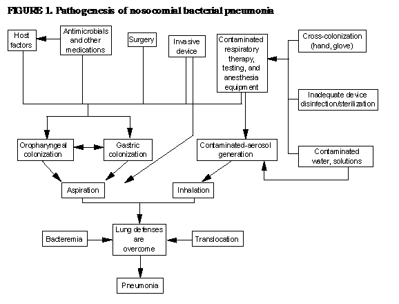