
Diagnosis of T. cruzi Reactivation in Immunosuppressed Patients
Diagnosis of T. cruzi reactivation requires demonstration of circulating parasite in peripheral blood or rising parasite numbers by quantitative (real time) PCR.
In organ recipients with skin lesions, T. cruzi may also be demonstrated in biopsy or aspirate.
In HIV/AIDS patients with central nervous system involvement, lesions may be visualized by MRI or other imaging techniques. T. cruzi may be demonstrable in CSF or biopsy by microscopy or molecular methods.
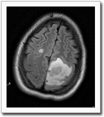
Magnetic resonance imaging (MRI) of the brain showing a mass lesion corresponding to a cerebral chagoma. Image courtesy of C.A. Arias, University of Texas, Houston.
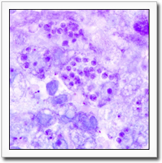
Nests of T. cruzi amastigotes in Giemsa-stained biopsy section from Chagas disease abscess in the brain of an HIV-coinfected patient (original magnification 1000X). Image courtesy of Dr. C.A. Arias, University of Texas, Houston.