
Case #145 - December, 2004
A 47-year-old woman traveled to the Dominican Republic. Upon her return she experienced fever and chills and went to see her doctor. A blood smear was ordered by her physician, stained with Wright’s-Giemsa, and examined. The following images show what was seen at 1000× magnification. What is your diagnosis? Based on what criteria?
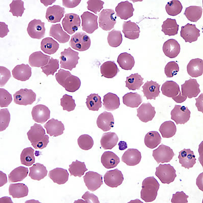
Figure A
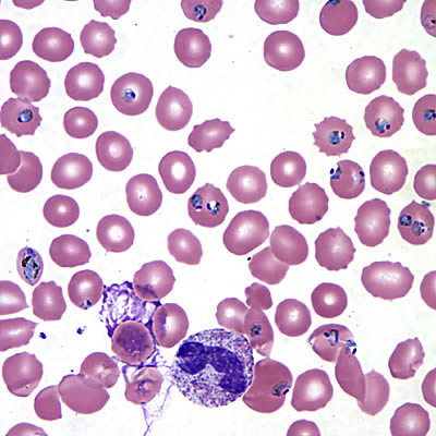
Figure B
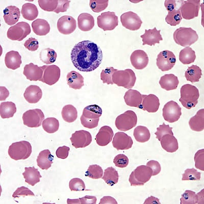
Figure C
Answer to Case #145
This was a case of malaria caused by Plasmodium falciparum. The morphology of the parasites was atypical, probably caused by a time lapse between blood collection and preparation of smears. The atypical presentation can be misleading, but key features can still be observed to allow a correct species identification. Morphologic features observed were:
- multiply infected RBC’s, which may indicate P. falciparum (although this can occur with other Plasmodium species).
- infected RBC’s that were normal-sized. Some of the uninfected, as well as the infected, RBC’s are crenated or irregular; no classic fimbriation was observed.
- appliqué forms.
- presence of mature trophozoites with pigment.
- presence of immature gametocytes (Figures B and C).
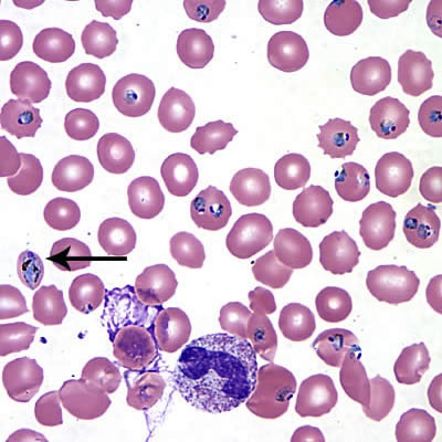
Figure A
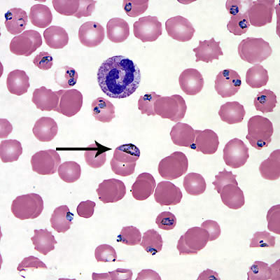
Figure B
More on: Malaria
This case was kindly contributed by the Illinois Department of Public Health Chicago Laboratory.
Images presented in the monthly case studies are from specimens submitted for diagnosis or archiving. On rare occasions, clinical histories given may be partly fictitious.
DPDx is an educational resource designed for health professionals and laboratory scientists. For an overview including prevention, control, and treatment visit www.cdc.gov/parasites/.