Case #366 – February 2014
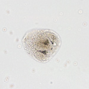
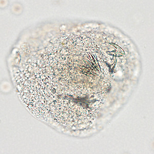
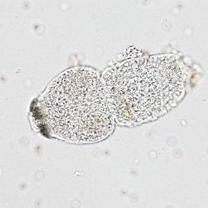
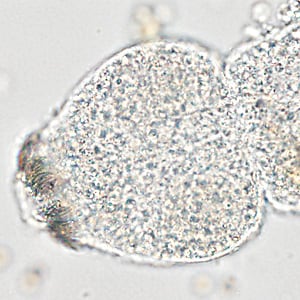
A 28-year-old male from Algeria had complaints of right upper quadrant (RUQ) abdominal pain. As part of his work up at a medical facility, imaging studies revealed a complex solid and cystic lesion in the posterior right hepatic dome. Hepatic cyst fluid was aspirated and submitted for ova-and-parasite (O&P) testing. Figures A–D show what was observed on a wet-mount made from the sediment of the fluid. Figures A and C were captured at 100x magnifications; Figures B and D at 200x magnification. What is your diagnosis? Based on what criteria?

Figure A

Figure B

Figure C

Figure D
Images presented in the DPDx case studies are from specimens submitted for diagnosis or archiving. On rare occasions, clinical histories given may be partly fictitious.
DPDx is an educational resource designed for health professionals and laboratory scientists. For an overview including prevention, control, and treatment visit www.cdc.gov/parasites/.