
Case #355 – September, 2013
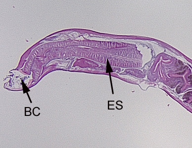
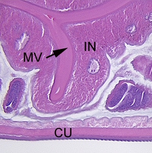
A worm-like object was removed during a routine colonoscopy performed on a 65-year-old male patient. The patient had no gastrointestinal complaints and no documented international travel. The object measured approximately 11.0 mm in length. The specimen was sent to Pathology for routine histological work-up, including sectioning and staining with hematoxylin-and-eosin (H&E). Digital images were captured and sent to the DPDx Team for diagnostic assistance. Figures A–D show four of the images sent for consultation. Follow-up ova-and-parasite (O&P) examinations of stool were negative. What is your diagnosis? Based on what criteria?

Figure A

Figure B

Figure C

Figure D
This case and images were kindly provided by Jackson Memorial Hospital, Miami, FL.
Images presented in the DPDx case studies are from specimens submitted for diagnosis or archiving. On rare occasions, clinical histories given may be partly fictitious.
DPDx is an educational resource designed for health professionals and laboratory scientists. For an overview including prevention, control, and treatment visit www.cdc.gov/parasites/.