
Case #326 – June, 2012
A 65-year-old female, who lives in northern California, had a colonoscopy performed as part of a routine screening. A foreign body was observed and subsequently removed. The specimen was sectioned, stained with hematoxylin and eosin (H&E), and examined by the attending pathologist. Digital images were captured and sent to the DPDx Team for diagnostic assistance. Figure A was captured at 2x; Figure B at 20x; and Figures C and D at 40x magnification. What is your diagnosis? Based on what criteria?

Figure A

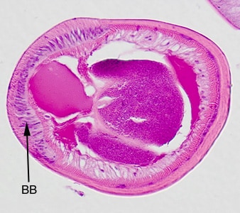
Figure B

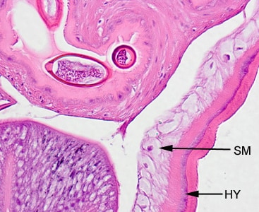
Figure C

Figure D
This case and images were kindly provided by Miraca Life Sciences, Phoenix, Arizona.
Images presented in the DPDx case studies are from specimens submitted for diagnosis or archiving. On rare occasions, clinical histories given may be partly fictitious.
DPDx is an educational resource designed for health professionals and laboratory scientists. For an overview including prevention, control, and treatment visit www.cdc.gov/parasites/.