
Case #317 – February, 2012
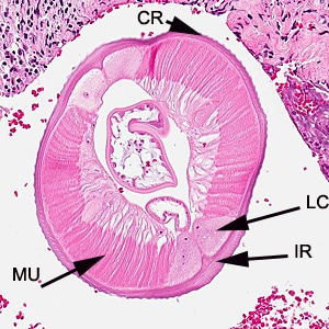
A 55-year-old woman sought medical attention for a nodule on the right side of her head. The patient presented with pain on the right side of her head for the past two months. Two weeks prior to seeking medical attention, she felt a painful 0.5 cm lump on her right temple. The nodule was removed surgically and sent to Pathology for histological testing. Stool and blood specimens were also collected, processed, and examined for parasites with negative results. The nodule was sectioned, stained with hematoxylin and eosin (H&E), and examined microscopically. Images were captured and sent to DPDx for diagnostic assistance. Figure A was captured at 40x; Figure B at 100x; Figures C and D at 200x magnification. What is your diagnosis? Based on what criteria?

Figure A

Figure B

Figure C

Figure D
This case and images were kindly provided by the National Public Health Surveillance Laboratory, Vilnius, Lithuania.
Images presented in the DPDx case studies are from specimens submitted for diagnosis or archiving. On rare occasions, clinical histories given may be partly fictitious.
DPDx is an educational resource designed for health professionals and laboratory scientists. For an overview including prevention, control, and treatment visit www.cdc.gov/parasites/.