
Case #308 – September, 2011
A 27-year-old patient underwent chemotherapy and a bone marrow transplant after being diagnosed with leukemia. Approximately two months after the bone marrow transplant, the patient was admitted to the hospital. The patient presented with severe, intractable cardiac decompensation and died while at the hospital. Autopsy specimens of the heart were sent to Pathology for routine sectioning and staining with hematoxylin and eosin (H&E). Figures A–D show what was observed by the attending pathologist on H&E-stained heart tissue. The slides and a tissue block were forwarded to the CDC’s Infectious Diseases Pathology Branch (IDPB) for further assistance. Specimens taken from the tissue block were also processed and examined by electron microscopy (Figures E and F) by IDPB and the DPDx Team. What is your diagnosis? Based on what criteria?

Figure A

Figure B

Figure C

Figure D

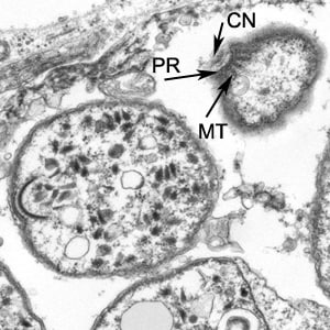
Figure E

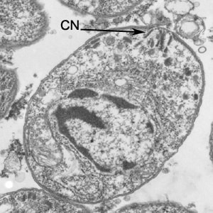
Figure F
Images presented in the DPDx case studies are from specimens submitted for diagnosis or archiving. On rare occasions, clinical histories given may be partly fictitious.
DPDx is an educational resource designed for health professionals and laboratory scientists. For an overview including prevention, control, and treatment visit www.cdc.gov/parasites/.