
Case #263 – November, 2009
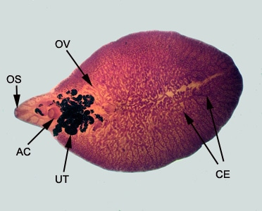
A 35-year-old man from Nepal presented at a hospital with recurring cholangitis; the patient had been experiencing symptoms for the previous six months. An MRI revealed two lesions in the liver, measuring approximately 5 centimeters long by 2 centimeters wide. Both lesions had cystic as well as solid components. An endoscopic retrograde cholangiopancreatography (ERCP) was also performed in the common bile duct, revealing flat leaf-shaped objects (Figures A–B). The objects were removed and collected in 10% formalin; they measured on average two centimeters in length (Figure C). One of the specimens was sent to a reference laboratory for identification, where it was stained with carmine (Figure D). What is your diagnosis? Based on what criteria?

Figure A

Figure B

Figure C

Figure D
This case and ERCP images were kindly provided by Dr. Subhash Agal, Kokilaben Dhirubhai Ambani Hospital, Mumbai, India.
Images presented in the DPDx case studies are from specimens submitted for diagnosis or archiving. On rare occasions, clinical histories given may be partly fictitious.
DPDx is an educational resource designed for health professionals and laboratory scientists. For an overview including prevention, control, and treatment visit www.cdc.gov/parasites/.