
Case #240 – November, 2008
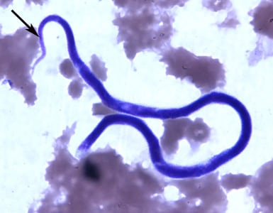
A 45-year-old immigrant from Mexico was admitted to the hospital after experiencing headaches, fever, pulmonary symptoms, and adenopathy. Because of his travel history, a blood specimen was collected and sent to Hematology for routine work-up, including blood parasitology. Figures A and B show objects observed on a Giemsa-stained thick smear of the blood specimen. Figures C and D show objects observed on a Giemsa-stained thin smear of the blood specimen. All images were captured at 1000x magnification. The objects in the figures measured on average 180 micrometers in length. What is your diagnosis? Based on what criteria?

Figure A

Figure B

Figure C

Figure D
Images presented in the DPDx case studies are from specimens submitted for diagnosis or archiving. On rare occasions, clinical histories given may be partly fictitious.
DPDx is an educational resource designed for health professionals and laboratory scientists. For an overview including prevention, control, and treatment visit www.cdc.gov/parasites/.