
Case #201 – April, 2007
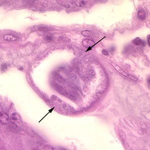
A 49-year-old Filipino man had an intestinal tissue biopsy taken. Figures A-C show what was seen on slides stained with hematoxylin and eosin (H & E). Figure A was taken at 100× magnification, Figure B was taken at 400× magnification, and Figure C (of a different section) was taken at 1000× magnification. Figure D was taken from a wet mount slide of formalin preserved stool; the object of interest in Figure D is approximately 40 micrometers long by 20 micrometers wide. What is your diagnosis? Based on what criteria?

Figure A

Figure B

Figure C

Figure D
Case study images were captured from reference material donated by Dr. John H. Cross of the Uniformed Services University of the Health Sciences, Bethesda, MD.
Images presented in the DPDx case studies are from specimens submitted for diagnosis or archiving. On rare occasions, clinical histories given may be partly fictitious.
DPDx is an educational resource designed for health professionals and laboratory scientists. For an overview including prevention, control, and treatment visit www.cdc.gov/parasites/.