
Case #125 - February, 2004
A seven-year-old child was taken to the doctor for abdominal pain, gas, bloating and intermittent diarrhea. The symptoms started about two days after returning from summer camp. Stool specimens were collected in formalin and polyvinyl alcohol (PVA) and sent to a commercial lab for ova-and-parasite (O&P) examination. Figures A–F show what was observed on a trichrome-stained smear made from the PVA-preserved stool. The images were captured at 1000x magnification. What is your diagnosis? Based on what criteria?
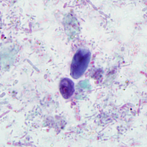
Figure A
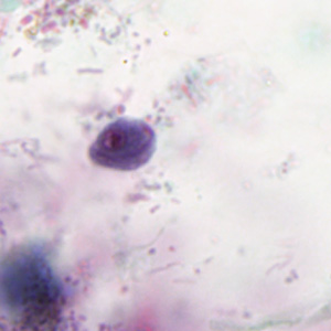
Figure B
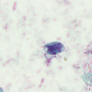
Figure C
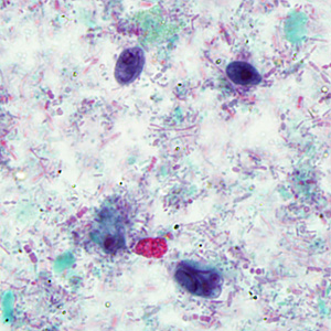
Figure D
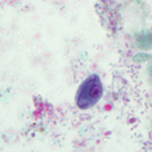
Figure E
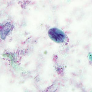
Figure F
This was case of giardiasis caused by Giardia duodenalis. Also present was Chilomastix mesnili, a flagellate generally considered nonpathogenic. Diagnostic morphologic features included:
- ovoid cysts of G. duodenalis (Figures C, F, and red arrows, Figures A and D), showing nuclei, axonemes and median bodies.
- oval to lemon-shaped cysts of C. mesnili (Figures B, E, and blue arrows, Figures A and D), showing a single nucleus and a cytostome running along a lateral edge. The Chilomastix cysts are slightly smaller than the Giardia cysts.

Figure A
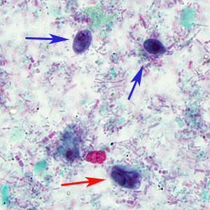
Figure D
More on: Giardiasis: Chilomastix mesnili
Images presented in the monthly case studies are from specimens submitted for diagnosis or archiving. On rare occasions, clinical histories given may be partly fictitious.
DPDx is an educational resource designed for health professionals and laboratory scientists. For an overview including prevention, control, and treatment visit www.cdc.gov/parasites/.