 ShareCompartir

Monthy Case Studies - 2003
Case #122 - December, 2003
A 24-year-old man experienced diarrhea that was mucoid and bloody, and he also had fever and gastrointestinal pain and cramping. His condition became severe and he was taken to the emergency room where physicians removed part of his colon. Figures A and B below show what was seen on a histological section from the colon, stained with hematoxylin and eosin (H & E). What is your diagnosis? Based on what criteria?
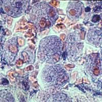
Figure A
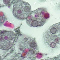
Figure B
Acknowledgement: This case kindly provided by the Frank Meglio from the Rhode Island Department of Health Laboratory and images were scanned from 2x2 Kodachrome slides.
Answer to Case #122
This was a case of invasive amebiasis caused by Entamoeba histolytica. Diagnostic features observed to identify trophozoites of E. histolytica included:
- trophozoites with a single nucleus containing even peripheral chromatin and ingested red blood cells.
- a size range consistent with E. histolytica versus white blood cells (macrophage ratio = 1:4-1:6; E. histolytica ratio = 1:10-1:20).
Serology is a useful tool to aid in the diagnosis of invasive or extraintestinal amebiasis prior to the use of invasive techniques. Commercial diagnostic laboratories offer a sensitive and specific enzyme immunoassay (EIA) test for the detection of antibodies to E. histolytica. In addition, some reference diagnostic laboratories can provide PCR for specific identification of E. histolytica.
More on: Amebiasis
Images presented in the monthly case studies are from specimens submitted for diagnosis or archiving. On rare occasions, clinical histories given may be partly fictitious.
