 ShareCompartir

Monthy Case Studies - 2003
Case #121 - December, 2003
A 70-year-old man was seen by an ophthalmologist for eye pain. Upon examination, the doctor found and removed a subconjuctival worm that was enclosed in a cyst. The man had no recent travel outside the United States (had traveled to Central America over 5 years ago) and reported that he did have pets at home. Figure A shows the cyst and the worm it contained; the worm was approximately 92 mm in length. Figure B shows the posterior end of the worm at 100× and Figure C shows the midbody of the worm at 200×. What is your diagnosis? Based on what criteria?
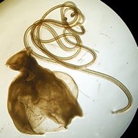
Figure A
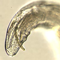
Figure B
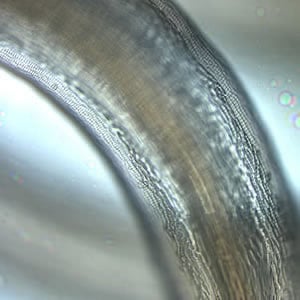
Figure C
Acknowledgement: This case kindly provided by the Dr. William Lushbaugh and the University of Mississippi Medical Center.
Answer to Case #121
This was a case of dirofilariasis, caused by a male filarial worm in the genus Dirofilaria. Based on absence of travel history and given the geographical location of the patient's residence, this worm is most likely Dirofilaria tenuis. The presentation of the worm in a cyst is not typical, but the morphology of the worm was consistent with the features of Dirofilaria. Diagnostic features observed included:
- cuticular ridging (Figuree A) that appeared beaded, in a "corn row" effect (blue arrows), indicating that the worm belonged to the genus Dirofilaria.
- caudal (tail) papillae (green arrow, Figure B).
- presence of a spicule (red arrow, Figure B), indicating this was a male worm.
- a size range compatible with adult Dirofilaria spp.
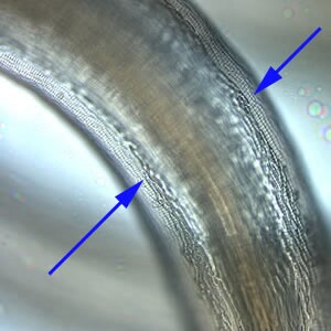
Figure C
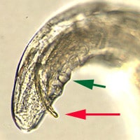
Figure B
Dirofilaria tenuis commonly infects raccoons, especially in the Southeastern United States, and zoonotic transmission can occur when mosquitoes feed on raccoons and subsequently on humans; human infection is considered accidental. Most cases in the United States of zoonotic Dirofilaria infections occurring in the subcutaneous tissues and eyes are caused by D. tenuis; D. immitis causes lung lesions.
More on: Dirofilariasis
Images presented in the monthly case studies are from specimens submitted for diagnosis or archiving. On rare occasions, clinical histories given may be partly fictitious.
