 ShareCompartir

Monthy Case Studies - 2003
Case #104 - March, 2003
A 38-year-old physician (radiologist) moved to the United States from India seven years ago, but he still makes frequent trips back to India. He noticed some enlargement of a mass in his shoulder (a subclavian mass) that had been there for three years. His physician performed a biopsy of the mass. Below are images from hematoxylin and eosin (H & E) stained section of the biopsied mass. What is your diagnosis? Based on what criteria?
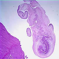
Figure A
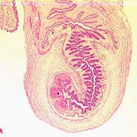
Figure B
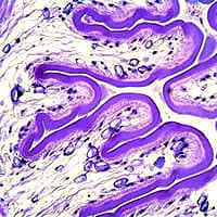
Figure C
Answer to Case #104
This was a case of cysticercosis caused by Taenia solium. Presence of cysticerci in subcutaneous tissue is a typical presentation of cysticercosis. Diagnostic features observed included:
- the cestode’s 'neck' region with part of an invaginated scolex (Figures A and C).
- the rostellar tissue and the suckers (Figure B, blue and black arrows, respectively).
- the presence of calcareous corpuscles (Figure C, black arrows).
- syncytial tegument and a parenchymous meshwork of internal tissue, both of which are characteristic of flatworms (trematodes and cestodes).
Figure A
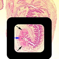
Figure B
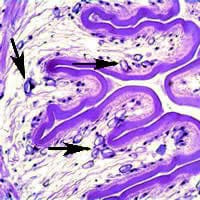
Figure C
More on: Cysticercosis
Images presented in the monthly case studies are from specimens submitted for diagnosis or archiving. On rare occasions, clinical histories given may be partly fictitious.
