 ShareCompartir

Monthy Case Studies - 2002
Case #97 - December, 2002
A 30-year-old woman traveled to Honduras last year. She did not take malaria prophylaxis. Since returning from her trip, she has experienced symptoms that included fever, chills, and headache. A blood sample was collected, smears were made and stained with Giemsa, and sent to the Tennessee Department of Health Laboratory Services. Below are images from her blood smears. What is your diagnosis? Based on what criteria?

Figure A
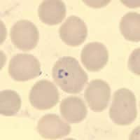
Figure B
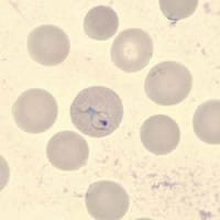
Figure C
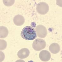
Figure D
Acknowledgement: The case history and images were kindly provided by the Tennessee Department of Health Laboratory Services.
Answer to Case #97
This was a case of malaria caused by Plasmodium vivax. Diagnostic features observed included:
Thick smear (Figure A):
- many parasites seen (rings, trophozoites, early schizonts, and gametocytes). The presence of many nuclei in the schizont was suggestive of P. vivax or P. falciparum; the morphology of the gametocytes helped rule out the latter.
Thin smear:
- a large, sturdy ring with a single chromatin dot (Figure B).
- an ameboid trophozoite in an enlarged red blood cell (1¼ to 1½ × normal size) with Schüffner's dots (Figure C).
- a large gametocyte, nearly 1½ to 2 × the normal size of a red blood cell, also with Schüffner's dots (Figure D).
More on: Malaria
Images presented in the monthly case studies are from specimens submitted for diagnosis or archiving. On rare occasions, clinical histories given may be partly fictitious.
