 ShareCompartir

Monthy Case Studies - 2002
Case #80 - March, 2002
A trichrome stained fecal smear was prepared and examined from the stool specimen of a 45-year-old serviceman returning from a tour of duty in the South Pacific. The objects observed in the smear ranged in size from 6 to 11 micrometers in diameter and are shown below (Figures A through F). What is your diagnosis? Based on what criteria?
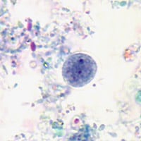
Figure A
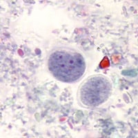
Figure B
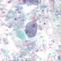
Figure C
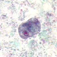
Figure D
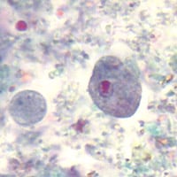
Figure E
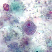
Figure F
Answer to Case #80
The objects shown in this case were Endolimax nana, a nonpathogenic ameba. Both cysts (Figures A and B) and trophozoites (Figures C through F) were shown in the case study. Diagnostic morphologic features observed included:
- the size of the organisms, which was consistent with cysts and trophozoites of E. nana.
- the presence of large karyosomes in the trophozoites, often appearing "blot-like" or irregularly shaped.
- coarse and vacuolated cytoplasm in some of the trophozoites.
- multinucleate cysts (which ruled-out the similar, Iodamoeba beutschlii, whose cyst stage has only one nucleus).
More on: Endolimax nana
Images presented in the monthly case studies are from specimens submitted for diagnosis or archiving. On rare occasions, clinical histories given may be partly fictitious.
