 ShareCompartir

Monthy Case Studies - 2002
Case #77 - February, 2002
A 4-year-old boy was seen in a children's hospital for fever, nausea, intermittent vomiting, anorexia, and diarrhea. Five months earlier, the boy had traveled to South America with his parents. The pediatrician ordered a complete blood count (CBC) and an O&P (ova and parasites) stool examination. The results from the blood count indicated eosinophilia. Three stool specimens from the boy were collected and preserved in formalin and PVA. The images are trichrome stained smears from his stool specimens (Figures A and B). The objects seen in the smears measured approximately 7 to 10 micrometers in length. What is your diagnosis? Based on what criteria?
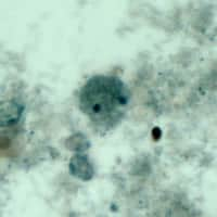
Figure A
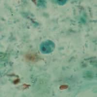
Figure B
Answer to Case #77
This was a case of intestinal infection due to Dientamoeba fragilis. The pathogenic status of D. fragilis is not well defined. However, cases have been reported in which D. fragilis is the only parasite found in stool specimens from children with symptoms of intestinal infection. Occasionally, children infected by this protozoan parasite present with eosinophilia. Diagnostic morphologic features included:
- the size range of the trophozoites, which was consistent with D. fragilis (5 to 15 micrometers).
- the presence of binucleate and uninucleate trophozoites (Figures A and B, respectively). Approximately 30 to 40% are uninucleate in most of the specimens.
- the absence of peripheral chromatin on the nuclear membrane.
- finely to heavily granular cytoplasm, which may be vacuolated, containing bacteria, yeast, and other debris.
More on: Dientamoeba fragilis
Images presented in the monthly case studies are from specimens submitted for diagnosis or archiving. On rare occasions, clinical histories given may be partly fictitious.
