 ShareCompartir

Monthy Case Studies - 2001
Case #70 - October, 2001
A woman submitted a stool specimen as part of her refugee screening. The specimen was subsequently divided into two aliquots: one was preserved in 10% formalin and the other in potassium dichromate. The specimens were submitted to the Florida Department of Health, Bureau of Laboratories. Microscopic examination revealed round objects measuring 10 to 12 µm in diameter; the laboratorians suspected Entamoeba histolytica and requested PCR. The objects are shown in Figures A, B, C, D, and E. What is your diagnosis? Based on what criteria?
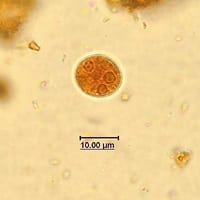
Figure A
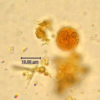
Figure B
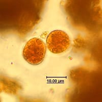
Figure C
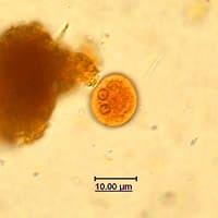
Figure D
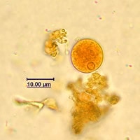
Figure E
Answer to Case #70
This case showed cysts and trophozoites of Entamoeba dispar. Based on the images presented in this case study, the correct answer was Entamoaeba histolytica/dispar because E. histolytica and E. dispar cannot be distinguished based on morphologic characteristics alone, unless trophozoites with ingested red blood cells are observed. Other tools are needed to differentiate the two species. PCR performed at the CDC identified the parasite as Entamoeba dispar. Diagnostic morphologic features included:
- a size range consistent with that of Entamoeba histolytica/dispar (10 to 20 micrometers; median range of 10 to 15 micrometers). Entamoeba hartmanni cysts resemble those of E. histolytica, however the size range is five to ten micrometers with a median range of six to eight micrometers.
- no more than four nuclei present in mature cysts.
- peripheral chromatin of the nuclei (cysts and trophozoites shown) that was heavy and evenly distributed.
More on: Intestinal Amebae
Images presented in the monthly case studies are from specimens submitted for diagnosis or archiving. On rare occasions, clinical histories given may be partly fictitious.
