 ShareCompartir

Monthy Case Studies - 2001
Case #69 - October, 2001
An adult man from Tacoma, WA developed fever, chills, nausea, arthralgias, and lethargy about two weeks after receiving a blood transfusion. Routine blood smears were prepared, stained with Giemsa, and examined in the hospital laboratory. Below are four images (Figures A, B, C, and D) taken from his blood smears. What is your diagnosis? Based on what criteria?
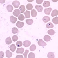
Figure A
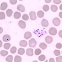
Figure B
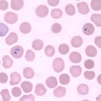
Figure C
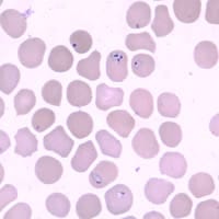
Figure D
Answer to Case #69
This was a case of babesiosis. Based on the patient's geographical location one might suspect the WA1 type that has been found in patients from the western United States. However, PCR identified the parasite as Babesia microti, not the WA1 type. Since the infected blood donor had not been identified when this case study was composed, we did not know where that person had acquired the infection. Diagnostic morphologic features included:
- pleomorphic ring-like parasites.
- dividing forms (Figures A, C, and D).
- an absence of pigment, even in the larger/older parasites.
- the presence of extraerythrocytic parasites were present (Figure B). This image was not a Plasmodium sp. schizont because there was no visible pigment. The argument could be raised that it might be a ruptured schizont, which could explain the lack of pigment, but the other morphological features of this case should be sufficient to rule out that possibility.
More on: Babesiosis
Images presented in the monthly case studies are from specimens submitted for diagnosis or archiving. On rare occasions, clinical histories given may be partly fictitious.
