 ShareCompartir

Monthy Case Studies - 2001
Case #59 - May, 2001
A 56-year-old man was admitted to a hospital in Arizona. He, his wife, and his sister-in-law had visited Tanzania and Kenya in recent weeks. He claimed that he had been bitten by insects (possibly biting flies), under the jaw, on the shoulder blades, and possibly on the lips. Approximately ten days after some of the bites, he became ill, complaining of fever, chills, headaches, back pain and malaise. He also had occasional bouts of nausea and vomiting and occasional right scotomata (dimness or dark spots in field of vision). His sister-in-law also received bites, but was asymptomatic. He visited a physician while overseas who prescribed Ciprofloxicin and doxycycline. At follow-up in Arizona, he had right submandibular swelling with superficial ulcerations with some serous drainage. There was also a quarter-sized induration/ulcer on his left scapula. The organisms shown in the following images were seen in his blood smears. What is your diagnosis? Based on what criteria?
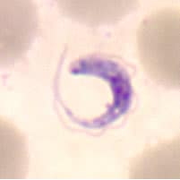
Figure A
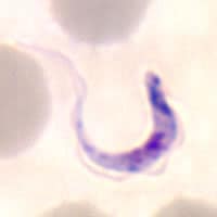
Figure B
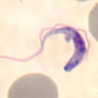
Figure C
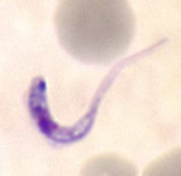
Figure D
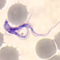
Figure E
Acknowledgement: This case was kindly provided by the Arizona State Laboratory, Infectious Disease Microbiology Section.
Answer to Case #59
This was a case of African trypanosomiasis probably caused by Trypanosoma brucei rhodesiense, based on epidemiological data that included the onset, severity of symptoms, and geographical location where the infection was acquired. Morphologically, the subspecies of African trypanosomes cannot be distinguished from one another. Diagnostic features observed were:
- organisms that exhibited a small kinetoplast. Trypanosoma cruzi trypomastigotes generally have a very large kinetoplast which may appear to bulge beyond the body of the organism. Even though some of the organisms shown have the characteristic "C" shape usually seen in T. cruzi trypomastigotes, the kinetoplasts are quite small. This, and the fact that there was no travel to Central or South America, should be adequate to rule out Chagas disease.
- the presence of dividing forms (Figure E), a feature seen in African trypanosomes and not in T. cruzi.
- the size of the organisms (average length about 20 micrometers), which helps to distinguish African trypanosomes from trypomastigotes of T. rangeli, which also have a small kinetoplast, but are longer and generally more slender (25 to 37 micrometers).
More on: African trypanosomiasis
Images presented in the monthly case studies are from specimens submitted for diagnosis or archiving. On rare occasions, clinical histories given may be partly fictitious.
