 ShareCompartir

Monthy Case Studies - 2001
Case #55 - March, 2001
A 33-year-old man returned to the United States after spending four years in Africa, mostly Kenya, working as an agricultural consultant. He had several parasitic diseases during his four years in Africa. After returning to the United States, he noticed a painful, swollen lesion with a black center on the top of his right foot. Initial diagnosis from a health care provider was that the lesion was most likely a plantar wart. The man did not agree with the physician and obtained a second opinion from a health care provider who suggested that the lesion be biopsied. The physician surgically removed the affected area and examined the excised tissue. The physician saw what appeared to be eggs in the tissue. The entire specimen was sent CDC's reference laboratory for identification of the eggs. The man did not have any complications from the surgery and the area healed within a few days. Figure A shows the entire specimen; all other images were taken from the cut surface of the tissue. Figures B and C were taken with a dissecting microscope equipped with adjustable zoom magnification. Figure D was taken with a compound microscope using a 5× objective and a fiber-optic light source to provide reflected light. What is your diagnosis? Based on what criteria?

Figure A
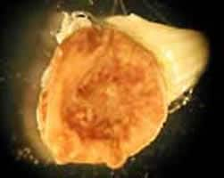
Figure B
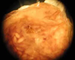
Figure C
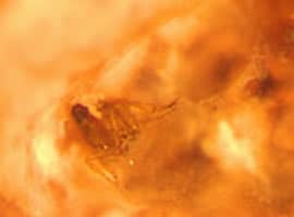
Figure D
Answer to Case #55
This was a case of tungiasis caused by Tunga penetrans, sometimes called a chigoe flea or jigger flea. The legs and eggs of the flea can be seen embedded in tissue in Figures B, C, and D. The inseminated female flea is about one millimeter in length. She burrows into the skin of the feet or lower legs, often on or between the toes, and feeds on blood. In the skin, the flea is oriented with her head facing away from the skin surface and her posterior end is sometimes visible as the characteristic black dot in the center of the lesion. The flea both breathes and discharges waste and eggs from the posterior end. After two weeks, the gravid female flea grows to a length of three to five millimeters and begins to discharge eggs. The eggs hatch and the larvae develop into adults in about three weeks. After copulation, the female flea searches for a warm-blooded mammalian host to which it will attach. If it does not find a suitable host it will die.
More on: Tungiasis
Images presented in the monthly case studies are from specimens submitted for diagnosis or archiving. On rare occasions, clinical histories given may be partly fictitious.
