 ShareCompartir

Monthy Case Studies - 2000
Case #41 - August, 2000
A 9-year-old boy was taken to his pediatrician for sudden headaches, fatigue, and loss of muscle coordination. His family lives on a ranch located approximately 80 miles northeast of San Francisco, CA. Clinical laboratory tests indicated eosinophilia and eosinophilic meningoencephalitis. Repeated stool tests were all negative for parasites. Significant in the history provided by the family was the fact that a family of raccoons had taken up residence in a storage shed in the same area of the yard where the child often plays. A brain biopsy was ordered and nematode larvae were found. Figure A shows what the pathologist found in an H & E (hemotoxylin and eosin) stained section obtained from a brain biopsy. An epidemiological investigation was initiated as a result of the nematode found in the biopsy. As part of this investigation, a pooled sample of raccoon feces was collected and examined for intestinal parasites. Numerous eggs similar to the one shown in the Figure B were seen. What is your diagnosis? Based on what criteria?
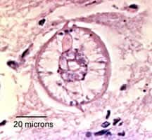
Figure A
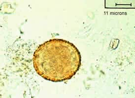
Figure B
Answer to Case #41
This was a case of baylisascariasis, caused by Baylisascaris, an intestinal ascarid of raccoons (B. procyonis) and skunks (B. columnaris). The image shows key diagnostic features of a typical ascarid larva, including prominent lateral alae, excretory columns (canals), and gut. The key morphological features for B. procyonis were:
- the size of the worm (about 50 micrometers in diameter), which was large enough to rule out Toxocara (typically 16 to 18 micrometers in diameter). The large size also excludes other larval nematodes such as Strongyloides and Trichinella. The morphology of these worms is also very different from ascarid larvae.
- the presence of two large, prominent lateral alae (LA, Figure A).
- the presence of large excretory columns (EC, Figure A) and a granular appearance of the intestines (Int, Figure A). The lumen (Lu, Figure A) is patent.
The size (about 80 by 70 micrometers) and morphology of the eggs found in the raccoon feces are also consistent with eggs of B. procyonis.
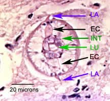
Figure A
More on: Baylisascariasis
Images presented in the monthly case studies are from specimens submitted for diagnosis or archiving. On rare occasions, clinical histories given may be partly fictitious.
