 ShareCompartir

Monthy Case Studies - 2000
Case #39 - July, 2000
An 42-year-old man with HIV was hospitalized for pneumonia. The attending physician ordered a bronchoalveolar lavage (BAL) and collected induced sputum specimens. The specimens were concentrated by centrifugation and smears were made from the sediment and stained with Giemsa. The objects shown below were seen on the slides (1000×, oil immersion). What is your diagnosis? Based on what criteria?
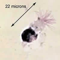
Figure A
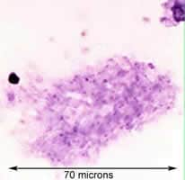
Figure B
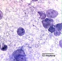
Figure C
Answer to Case #39
This was a case of Pneumocystis jiroveci pneumonia. Diagnostic features observed were:
- a mass of trophozoites of Pneumocystis jiroveci , also known as a syncytial mass (Figure B). You can see many nuclei within the mass of cytoplasm. This aggregation of trophozoites is generally only seen in samples obtained using invasive procedures such as a BAL. Often, only isolated or much smaller groups of trophozoites are seen and sometimes a few cysts can be detected as well.
- the cyst stage of Pneumocystis jiroveci (Figure C). The size of these structures (four to five micrometers) is consistent with the lower end of Pneumocystis jiroveci (four to eight micrometers) and you can see intracystic bodies (sometimes referred to as sporozoites) within the cysts. There can be up to eight such bodies within a cyst, each with a distinct nucleus. When stained with Giemsa, the actual cyst wall does not stain; however the intracystic bodies will stain and will appear to be in a uniform clear area on the smear. Instead of Giemsa, some laboratories use Gomori's methenamine silver stain, which will stain only the cyst wall. Indirect fluorescent antibody tests (IFA) and PCR methods are also used to diagnose an infection with Pneumocystis.
The object in Figure A was a ciliated epithelial cell, and not a parasite.
Note: Pneumocystis, at one time, was thought by some to be a protozoan while others believed that it was more closely related to a fungus. Molecular tools have shown in recent years that it is phylogenetically related to fungi (Edman JC, Kovacs JA, Masur H, Santi DV, Elwood HJ, Sogin ML. Ribosomal RNA sequence shows Pneumocystis jiroveci to be a member of the fungi. Nature 1988 Aug 11;334 (6182):519-22). Many parasitology books and atlases still refer to it as a protozoan.
More on: Pneumocystis
Images presented in the monthly case studies are from specimens submitted for diagnosis or archiving. On rare occasions, clinical histories given may be partly fictitious.
