 ShareCompartir

Monthy Case Studies - 2000
Case #32 - March, 2000
A 39-year-old middle school biology teacher collected shells as souvenirs for her students from her summer vacation in Thailand and the Philippines. She collected a large number of shells from beaches, lakes, rivers, and streams of both countries. The types of shells she collected are shown in Figures A and B. After returning from her trip, she went to a local health clinic for mild to moderate episodes of intestinal cramping. Within the last two to three weeks, she indicated that the cramping became more severe and frequent. She also stated that she had occasionally seen blood in her stool and that her abdomen was slightly bloated. She submitted a stool specimen for examination. An FEA concentration was performed on her stool and a wet mount of the concentrate was examined. Eosinophilia was noted in her bloodwork. The object in Figure C was seen in moderate numbers (three to nine objects per 22 mm coverslip at 100× magnification). The average size of the objects was 90 by 60 micrometers. What is your diagnosis? Based on what criteria?
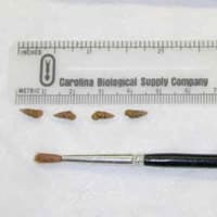
Figure A
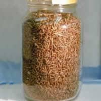
Figure B
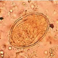
Figure C
Answer to Case #32
This was a case of schistosomiasis caused by Schistosoma japonicum. Diagnostic features observed were:
- an egg (Figure C) within the size range for S. japonicum (70 to 100 micrometers). S. mekongi eggs are generally smaller (51 to 78 micrometers).
- a lack of an operculum on the egg.
- the presence of a miracidium within the egg.
- the presence of a small, inconspicuous spine (though out of the focal plane in the image) on the egg.
Most of the respondents identified the snail shells as those of Oncomelania spp., the intermediate snail host for S. japonicum.
More on: Schistosomiasis
Images presented in the monthly case studies are from specimens submitted for diagnosis or archiving. On rare occasions, clinical histories given may be partly fictitious.
