 ShareCompartir

Monthy Case Studies - 2000
Case #29 - February, 2000
A 3-year-old girl was seen in a small rural clinic in the Southeastern United States with complaints of abdominal pain and upset stomach. A local laboratory performed a routine stool examination and observed the objects shown in the images below. The laboratory suspected the objects were artifacts. A portion of the stool concentrate was sent to CDC's reference laboratory for confirmation that parasite cysts or eggs were not present. What is your diagnosis? Based on what criteria?
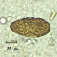
Figure A
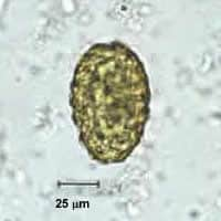
Figure B
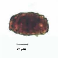
Figure C
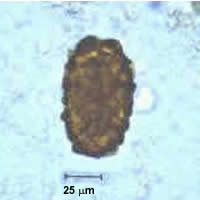
Figure D
Answer to Case #29
This was a case of ascariasis caused by Ascaris lumbricoides. The objects shown in the images were infertile Ascaris eggs. The image below (Figure E) shows infertile and fertile Ascaris eggs for comparative purposes. Infertile Ascaris eggs can be difficult to identify because of variations in size, shape, and surface of the egg shell. Infertile Ascaris eggs tend to be larger and more elongate than fertile Ascaris eggs and they measure approximately 80 to 95 micrometers long by 40 to 50 micrometers wide. The shell can either be mammillated, mammillated with a grossly distorted surface, or relatively smooth without mammillation (similar to decorticated fertile eggs). Infertile Ascaris eggs may be seen in early infections where the female worms have not yet mated, or in areas of low exposure where the chance of single sex infections is higher. The health concerns of an Ascaris infection are the same whether fertile or infertile eggs are passed, so treatment would be recommended.
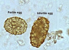
Figure E
More on: Ascariasis
Images presented in the monthly case studies are from specimens submitted for diagnosis or archiving. On rare occasions, clinical histories given may be partly fictitious.
