 ShareCompartir

Monthy Case Studies - 1999
Case #25 - December, 1999
The object in the following image was found in a wound on the toe of a field worker from Ecuador. The size of this object is 640 micrometers by 350 micrometers. What is your diagnosis? Based on what criteria?
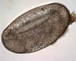
Figure A
Acknowledgement: This case and image were kindly provided by the Oregon Public Health Laboratory.
Answer to Case #25
This was a case of tungiasis caused by Tunga penetrans, a parasitic flea. The object in Figure A was an egg of T. penetrans. Adults of this species live in the sand and are found in tropical climates. Impregnated female fleas latch on to the feet or lower extremities of a host (human or animal). The fleas then invade the subcutaneous tissue where they feed, increase in size, produce eggs, and cause a small, ulcer-like lesion. The eggs generally fall off the host after being expelled by the female flea, which explains why you do not usually see eggs of T. penetrans in cases of tungiasis.
More on: Tungiasis
Images presented in the monthly case studies are from specimens submitted for diagnosis or archiving. On rare occasions, clinical histories given may be partly fictitious.
