 ShareCompartir

Monthy Case Studies - 1999
Case #15 - July, 1999
The following images are fecal smears (trichrome stained) from a 61-year-old man with bloody and mucoid diarrhea. He frequently traveled between Texas and Mexico. What is your diagnosis? Based on what criteria?
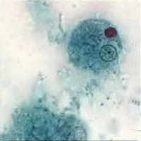
Figure A
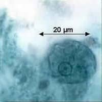
Figure B
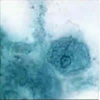
Figure C
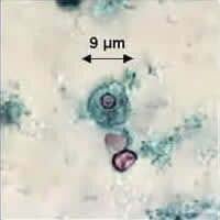
Figure D
Answer to Case #15
This was a case of amebiasis caused by Entamoeba histolytica. Figure A showed a trophozoite with a nucleus that is typical of E. histolytica/dispar with a small, central karyosome and fine, evenly distributed peripheral chromatin. That the infection is caused by E. histolytica instead of E. dispar is based on the fact that the trophozoite in Figure A has ingested an erythrocyte (appearing as a dark circle in the right upper quadrant). Infection by E. histolytica is also suggested by the history of bloody diarrhea.
Our colleagues from the Texas Department of Health sent us the smears because they also saw some parasites that looked like Entamoeba coli, and wanted a second opinion. Indeed, in the smear there are some parasites that look like E. coli. For example, the trophozoite in Figure C has a nucleus with peripheral chromatin that is somewhat coarse and uneven, suggesting E. coli. However, the specimen overall does not appear to have been very well preserved, and in that situation E. histolytica/dispar can somewhat resemble E. coli (as one of the responders elegantly put it: "irregular but not beyond the normal variance"). For additional certainty, we emailed images of the specimen, and then sent the smear itself, to Larry Ash (a member of the email network, in Los Angeles), who also thought that this was E. histolytica (technically, we could not do PCR for Entamoeba species on this permanently stained smear).
Finally, Figure D showed two erythrocytes and, above them, a leukocyte that should be differentiated from a small amebae.
More on: Amebiasis
Images presented in the monthly case studies are from specimens submitted for diagnosis or archiving. On rare occasions, clinical histories given may be partly fictitious.
