Isolation of Diverse Simian Arteriviruses Causing Hemorrhagic Disease
Teressa M. Shaw, Samuel T. Dettle, Andres Mejia, Jennifer M. Hayes, Heather A. Simmons, Puja Basu, Jens H. Kuhn, Mitchell D. Ramuta, Cody J. Warren, Peter B. Jahrling, David H. O’Connor, Liupei Huang, Misbah Zaeem, Jiwon Seo, Igor I. Slukvin, Matthew E. Brown, and Adam L. Bailey

Author affiliations: University of Wisconsin, Madison, Wisconsin, USA (T.M. Shaw, S.T. Dettle, A. Mejia, M.D. Ramuta, D.H. O’Connor, L. Huang, M. Zaeem, J. Seo, I.I. Slukvin, M.E. Brown, A.L. Bailey); Wisconsin National Primate Research Center, Madison (S.T. Dettle, A. Mejia, J.M. Hayes, H.A. Simmons, P. Basu, D.H. O’Connor, I.I. Slukvin); National Institutes of Health, Fort Detrick, Frederick, Maryland, USA (J.H. Kuhn, P.B. Jahrling); The Ohio State University, Columbus, Ohio, USA (C.J. Warren)
Main Article
Figure 5
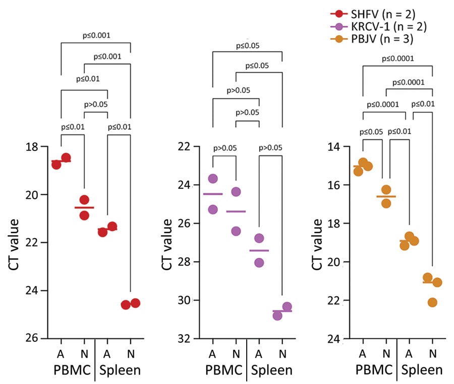
Figure 5. Comparisons of Ct values for different viruses infecting cells from rhesus monkeys in study of diverse simarteriviruses causing hemorrhagic disease. PBMCs and splenocytes were isolated from rhesus monkeys, infected with different simarteriviruses, and analyzed for infection by using quantitative reverse transcription PCR. Dots for each cell type and numbers in parentheses indicate number of experiments performed for each virus. Horizontal lines between dots indicate mean Ct values for each group. Statistical significance was determined by using 1-way analysis of variance. A, adherent; Ct, cycle threshold; KRCV-1, Kibale red colobus monkey virus 1; N, nonadherent; PBJV, Pebjah virus; PBMCs, peripheral blood mononuclear cells; SHFV, simian hemorrhagic fever virus.
Main Article
Page created: February 15, 2024
Page updated: March 20, 2024
Page reviewed: March 20, 2024
The conclusions, findings, and opinions expressed by authors contributing to this journal do not necessarily reflect the official position of the U.S. Department of Health and Human Services, the Public Health Service, the Centers for Disease Control and Prevention, or the authors' affiliated institutions. Use of trade names is for identification only and does not imply endorsement by any of the groups named above.