Isolation of Diverse Simian Arteriviruses Causing Hemorrhagic Disease
Teressa M. Shaw, Samuel T. Dettle, Andres Mejia, Jennifer M. Hayes, Heather A. Simmons, Puja Basu, Jens H. Kuhn, Mitchell D. Ramuta, Cody J. Warren, Peter B. Jahrling, David H. O’Connor, Liupei Huang, Misbah Zaeem, Jiwon Seo, Igor I. Slukvin, Matthew E. Brown, and Adam L. Bailey

Author affiliations: University of Wisconsin, Madison, Wisconsin, USA (T.M. Shaw, S.T. Dettle, A. Mejia, M.D. Ramuta, D.H. O’Connor, L. Huang, M. Zaeem, J. Seo, I.I. Slukvin, M.E. Brown, A.L. Bailey); Wisconsin National Primate Research Center, Madison (S.T. Dettle, A. Mejia, J.M. Hayes, H.A. Simmons, P. Basu, D.H. O’Connor, I.I. Slukvin); National Institutes of Health, Fort Detrick, Frederick, Maryland, USA (J.H. Kuhn, P.B. Jahrling); The Ohio State University, Columbus, Ohio, USA (C.J. Warren)
Main Article
Figure 4
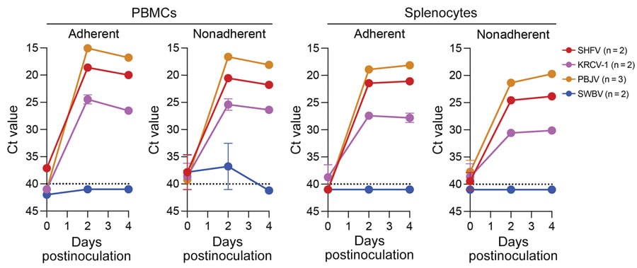
Figure 4. Ct values for virus-specific nucleoprotein RNA from rhesus monkey cells in study of diverse simarteriviruses causing hemorrhagic disease. PBMCs and splenocytes were infected with SHFV at a multiplicity of infection of 0.1 and infected with KRCV-1, PBJV, or SWBV-1 by using volumes equivalent to that of SHFV. Nucleoprotein RNA was measured by using quantitative reverse transcription PCR at different times after inoculation. Dotted lines indicate limit of detection. Numbers in parentheses indicate number of experiments performed for each virus. Error bars indicate SEMs. Ct, cycle threshold; KRCV-1, Kibale red colobus monkey virus 1; PBJV, Pebjah virus; PBMCs, peripheral blood mononuclear cells; SHFV, simian hemorrhagic fever virus; SWBV-1, Southwest baboon virus 1.
Main Article
Page created: February 15, 2024
Page updated: March 20, 2024
Page reviewed: March 20, 2024
The conclusions, findings, and opinions expressed by authors contributing to this journal do not necessarily reflect the official position of the U.S. Department of Health and Human Services, the Public Health Service, the Centers for Disease Control and Prevention, or the authors' affiliated institutions. Use of trade names is for identification only and does not imply endorsement by any of the groups named above.