Volume 30, Number 4—April 2024
Research
Isolation of Diverse Simian Arteriviruses Causing Hemorrhagic Disease
Teressa M. Shaw, Samuel T. Dettle, Andres Mejia, Jennifer M. Hayes, Heather A. Simmons, Puja Basu, Jens H. Kuhn, Mitchell D. Ramuta, Cody J. Warren, Peter B. Jahrling, David H. O’Connor, Liupei Huang, Misbah Zaeem, Jiwon Seo, Igor I. Slukvin, Matthew E. Brown, and Adam L. Bailey
Figure 2
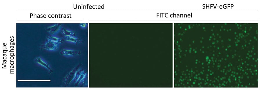
Figure 2. Primary rhesus monkey macrophages infected with simian hemorrhagic fever virus in study of diverse simarteriviruses causing hemorrhagic disease. Macrophages were isolated from spleen tissue and mock infected or infected with SHFV-eGFP. Left panel shows isolated macrophages; scale bar indicates 120 μm. Middle panel shows cells 24 hours after mock infection and right panel shows cells 24 hours after infection with SHFV-eGFP at a multiplicity of infection of 0.1; original magnification ×40. SHFV-eGFP, simian hemorrhagic fever virus–enhanced green fluorescent protein.
Page created: February 15, 2024
Page updated: March 20, 2024
Page reviewed: March 20, 2024
The conclusions, findings, and opinions expressed by authors contributing to this journal do not necessarily reflect the official position of the U.S. Department of Health and Human Services, the Public Health Service, the Centers for Disease Control and Prevention, or the authors' affiliated institutions. Use of trade names is for identification only and does not imply endorsement by any of the groups named above.