Meningococcal Photos
WARNING: Some of these photos might be unsuitable for children. Viewing discretion is advised.
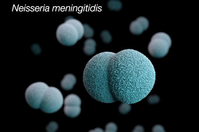
This illustration depicts a three-dimensional (3D) computer-generated image of a number of diplococcal, Gram-negative, Neisseria meningitidis, bacteria. The artistic recreation was based upon scanning electron microscopic (SEM) imagery.
Source: PHIL Photo ID# 22881
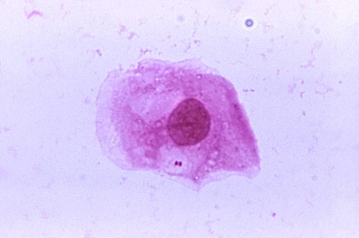
A photomicrograph of Neisseria meningitidis recovered from the urethra of an asymptomatic male; Magnified 1125X (N. meningitidis is responsible for causing “meningococcal” meningococcal. This bacterium is not normal flora, but a pathogenic organism that may be present in a large percentage of the population without causing disease).
Source: PHIL Photo ID# 2678
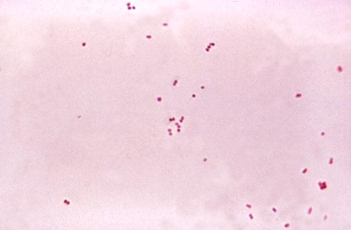
This micrograph depicts the presence of aerobic Gram-negative Neisseria meningitidis diplococcal bacteria; Mag. 1150X (Meningococcal disease is an infection caused by a bacterium called N. meningitidis or the meningococcus. The meningococcus lives in the throat of 5-10% of healthy people. Rarely, it can cause serious illness such as meningococcal or blood infection).
Source: PHIL Photo ID# 6423
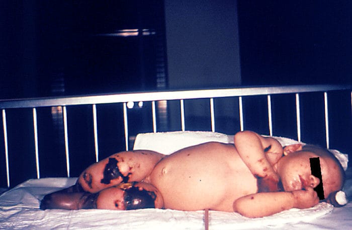
4 month old female with gangrene of hands and lower extremities due to meningococcemia.
Source: PHIL Photo ID#
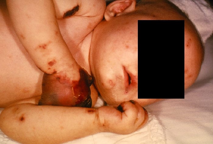
4 month old female with gangrene of hands due to meningococcemia.
Source: PHIL Photo ID#