Volume 15, Number 2—February 2009
Dispatch
Isolation of Kyasanur Forest Disease Virus from Febrile Patient, Yunnan, China
Abstract
We recently determined that Nanjianyin virus, isolated from serum of a patient in Yunnan Province, China, in 1989, is a type of Kyasanur Forest disease virus. Results of a 1987–1990 seroepidemiologic investigation in Yunnan Province had shown that residents of the Hengduan Mountain region had been infected with Nanjianyin virus.
Figure 1
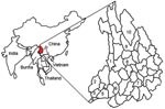
Figure 1. Counties in the Hengduan Mountain region of Yunnan Province where Kyasanur Forest disease virus antibody has been detected. 1, Lushuii County, antibody found in 31.6% of humans, 25.5% of birds, and...
Kyasanur Forest disease (KFD) virus, a member of the tick-borne encephalitis virus serocomplex of the genus Flavivirus, family Flaviviridae, can cause fever, hemorrhage, and encephalitis and has a 3%–5% case-fatality ratio (1). KFD was discovered in 1957 in the Mysore forest region of south India, where 400–500 persons per year were infected with the virus (2,3). KFD virus has been found only in monkeys, humans, and Haemaphysalis spinigera ticks in the KFD-epidemic region of south India (4), although a variant of KFD virus, Alkhurma virus, was isolated recently in Saudi Arabia (5). In this study, we determined that the gene sequence of a Nanjianyin virus isolate obtained from a febrile patient is highly homologous to that of KFD virus. The Nanjianyin virus was isolated in 1989 from the serum of a 38-year-old woman from the Hengduan Mountain region of Yunnan Province, People’s Republic of China, where a previous serosurvey demonstrated that KFD exposure had occurred (Figure 1).
In tests conducted shortly after isolation of Nanjianyin virus in 1989, the virus caused a typical cytopathic effect within 4 days after its injection in BHK-21 cells, killed 100% of 3-day-old mice within 2.5 days after their intracerebral inoculation with a 25-μL culture supernatant, and killed 100% of 50-day-old adult mice within 11–13 days of their intraperitoneal inoculation with a 30-μL culture supernatant. Hemagglutination inhibition test results showing a cross-reaction between Nanjinayin virus and a Japanese encephalitis virus antibody indicated that Nanjianyin virus belonged to the genus Flavivirus. No further tests to classify Nanjianyin virus were performed at the time it was isolated. The virus was preserved by lyophilization and stored at –30°C.
Recently, we used molecular methods to determine that Nanjianyin virus is a variant of KFD virus. After reconstituting the lyophilized virus in a BioSafety Level 3 biosafety cabinet, we suspended the sample in 0.5 mL minimum essential media (Gibcol BRL, Gaithersburg, MD, USA) (pH 7.4) and then centrifuged it for 5 min at 6,000× g. We then extracted the total RNA from 140 µL of supernatant by using the QIAamp Viral RNA Mini Kit (QIAGEN, Valencia, CA, USA) in accordance with the manufacturer’s protocol and produced the first strands of cDNA by using Ready-To-Go You-Prime First-Strand Beads (Amersham Pharmacia Biotech, Piscatawy, NJ, USA) as described in the manual accompanying the kit. We used Flavivirus genus-specific primers (6) to perform reverse transcription-PCR amplification using viral genomic RNA as a template and determined the nucleotide sequence of the virus from the amplified cDNA fragment. Results of nucleotide sequence analysis by BLAST (http://blast.ncbi.nlm.nih.gov/Blast.cgi) showed that the nucleotide DNA sequence of Nanjianyin virus was 99% homologous to that of KFD virus (prototype KFDV Itp9605, GenBank accession no. AY323490).
To complete the sequence determination of the PrM-E genes, we designed 3 pairs of primers to amplify them. Using information from a previous study (6), we also designed an additional primer pair to amplify the nonstructural protein (NS5) gene (Table).
Figure 2
Figure 2. Phylogenetic analysis of the PrM-E (A) and nonstructural protein 5 (B) gene sequences of Nanjianyin virus isolated from Yunnan Province, China. Phylogenetic analyses were performed by the neighbor-joining method with MEGA...
Results of sequence alignment and homology analysis performed with MegAlign software of DNASTAR (Madison, WI, USA) showed that the 654-bp PrM gene of Nanjianyin virus was 99.6% identical to that of KFD virus (Itp9605 strain), 99.4% identical to that of KFD virus (EU480489), 98.2% identical to that of KFD virus (X74111), but only 90.4% identical to that of Alkhurma hemorrhagic fever (AFH) virus (1176 strain), and only 57.2% to 64.3% identical to the 654-bp PrM genes of other tick-borne encephalitis complex viruses such as Omsk hemorrhagic fever virus (Kubrin strain), tick-borne encephalitis virus (Sengzhang strain), Powassan virus (LB strain), and Langat virus (TP21 strain). The 1487-bp E gene nucleotide sequence of Nanjianyin virus was 99.8% identical to that of KFD virus (Itp9605 strain), 99.8% identical to that of KFD virus (EU480489), 98.5.0% identical to that of KFD virus (X74111), 91.9% identical to that of AFH virus (1176 strain), and <72% identical to that of other tick-borne encephalitis complex viruses. The nucleotide sequence of the 1,000-bp NS5 gene of Nanjianyin virus was 99.6%, 99.7%, and 99.7% homologous to that of KFD virus (Itp9605 strain), KFD virus (W371), and KFD virus (EU480489), respectively; 92.3% homologous to that of AFH virus isolate 1176; and <77.6% homologous to the 1,000-bp NS5 gene of other tick-borne encephalitis complex viruses. Results of homology analyses thus demonstrated that Nanjianyin virus belongs to the KFD virus clade, and results of phylogenetic analyses conducted with 2,142 nt of the PrM-E gene and 1,000 nt of the NS5 gene suggested that Nanjianyin virus and KFD virus are in the same genetic cluster (Figure 2).
Results of a serosurvey of tick-borne viruses conducted from 1987 through 1990 in Yunnan Province (7) showed that 169 (19.5%) of 867 healthy residents of western Yunnan Province (in Lushui, Shidian, Yingjiang, Mangshi, Ruili, and Longchuan counties) and 6 (3.7%) of 161 healthy residents of northwestern Yunnan Province (in Lijiang and Diqin counties) carried antibodies against KFD virus. KFD antibodies also were detected in the serum of patients with fever in Lushui County (7,8) and in the serum of resident birds, migratory birds, rodents, and rhesus monkeys (Macaca mulatta) in the Hengduan Mountain region (Lushui and Eryuan counties) (7,9). These results indicate that humans and animals in the Hengduan Mountain region of Yunnan Province have been infected with KFD virus since the 1980s. Although detailed information about the movement of the woman infected with Nanjianyin virus in 1989 is not available, residents of the Hengduan Mountain region at that time seldom traveled far, so she probably was exposed there.
Results of epidemiologic and virologic investigations suggest that migratory birds play a key role in the spread of arboviruses (10,11). Migratory birds frequently pass through Yunnan Province during their migration from south India and the Indian Ocean islands to Mongolia and Siberia. The areas adjacent to Hengduan Mountain in Yunnan Province and India also provide a suitable habitat for Haemaphysalis spinigera, which is the vector for KFD virus in the region (12,13). Our results, combined with those in previous seroprevalence reports of KFD virus in humans and birds (6,7), indicate that KFD virus likely was carried to the region by these migratory birds and their parasitic ticks. KFD antibodies have been detected in residents of north and northeast India, and the KFD seropositive rate is especially high among residents of India’s Andaman Islands and Nicobar Islands (14). KFD antibodies also were detected in both human and bird serum in the Chinese districts of Guangdong, Guangxi, Guizhou, Hubei, Henan, Xinjiang, and Qinghai in 1983 (15).
In summary, we found that Nanjianyin virus, first isolated in the Hengduan Mountain region of Yunnan Province, is a variant of KFD virus. This finding confirms that infection with KFD virus has previously occurred in the region and justifies enhanced surveillance for KFD among febrile patients in the Hengduan Mountain region.
Mr Wang is a PhD candidate at the Institute for Viral Disease Control and Prevention, Chinese Center for Disease Control and Prevention. He specializes in medical microbiology, and his current research interests include the detection and diagnosis of emerging infectious agents.
Acknowledgment
This work was supported by grants from the Ministry of Science and Technology of China (no. 2003BA712A08-01); the National Natural Science Foundation of China (no. 30560142); and China CDC–US CDC Cooperative Agreement U19-GH000004.
References
- Lin D, Li L, Dick D, Shope RE, Feldmann H, Barrett ADT, Analysis of the complete genome of the tick-borne flavivirus Omsk hemorrhagic fever virus. Virology. 2003;313:81–90. DOIPubMedGoogle Scholar
- Work TH, Trapido H, Narasimha Murthy DP, Laxmana RR, Bhatt PN, Kulkarni KG. Kyasanur Forest disease III: a preliminary report on the nature of the infection and clinical manifestations in human beings. Indian J Med Sci. 1957;11:619–45.PubMedGoogle Scholar
- Banerjee K. Kyasanur Forest disease. In: Monath TP, editor. Arboviruses epidemiology and ecology. Boca Raton (FL): CRC Press; 1988. p. 93–116.
- Pattnaik P. Kyasanur Forest disease: an epidemiological view in India. Rev Med Virol. 2006;16:151–65. DOIPubMedGoogle Scholar
- Zaki AM. Isolation of a flavivirus related to the tick-borne encephalitis complex from human cases in Saudi Arabia. Trans R Soc Trop Med Hyg. 1997;91:179–81. DOIPubMedGoogle Scholar
- Kuno G, Chang GJ, Tsuchiya KR, Karabatsos N, Cropp CB. Phylogeny of the genus flavivirus. J Virol. 1998;72:73–83.PubMedGoogle Scholar
- Hou ZL, Huang WL, Zi DY, Zhang HL, Shi HF. Study of the serologic epidemiology of tick-borne viruses in Yunnan [in Chinese]. Chinese Journal of Vector Biology and Control. 1992;3:173–6.
- Zhang TS, Wang YM, Zhang YH, Duan S. A survey of antibodies to arboviruses in residents of southwestern Yunnan Province [in Chinese]. Chin J Endemiology. 1989;10:74–7.
- Yang QR, Liu XZ, Zhang JY, Zi DY, Zhang HL. A study of arbovirus antibodies in birds of the Niao-Diao mountain area of Eryuan County in Yunnan Province [in Chinese]. Chin J Endemiology. 1988;9:150–3.
- Ghosh SN, Rajagopalan PK, Singh GK, Bhat HR. Serological evidence of arbovirus activity in birds of KFD epizootic—epidemic area, Shimoga District, Karnataka, India. Indian J Med Res. 1975;63:1327–34.PubMedGoogle Scholar
- Venugopal K, Buckley A, Reid HW, Gould EA. Nucleotide sequence of the envelope glycoprotein of Negishi virus show close homology to louping ill virus. Virology. 1992;190:515–21. DOIPubMedGoogle Scholar
- Gong ZD, Hai BQ. Investigation of small animals in the Gaoli Mountain region [in Chinese]. Journal of Veterinary Medicine. 1989;24:28–32.
- Gong ZD, Zi DY, Feng XG. Composition and distribute of ticks in the Hengduan Mountain region of western Yunnan, China [in Chinese]. Chinese Journal of Pest Control. 2001;2:13–5.
- Padbidri VS, Wairagkar NS, Joshi GD, Umarani UB, Risbud AR, Gaikwad DL, A serological survey of arboviral diseases among the human population of the Andaman and Nicobar Islands, India. Southeast Asian J Trop Med Public Health. 2002;33:794–800.PubMedGoogle Scholar
- Chen BQ, Liu QZ, Zhou GF. Investigation of arbovirus antibodies in serum from residents of certain areas of China [in Chinese]. Chin J Endemiology. 1983;4:263–6.
Figures
Table
Cite This ArticleTable of Contents – Volume 15, Number 2—February 2009
| EID Search Options |
|---|
|
|
|
|
|
|
Please use the form below to submit correspondence to the authors or contact them at the following address:
Guo-Dong Liang, State Key Laboratory for Infectious Disease Prevention and Control, Institute for Viral Disease Control and Prevention, Chinese Center for Disease Control and Prevention, 100 Yingxin Street, Xuanwu District, Beijing 100052, China
Top