bannermenu

Trichostrongylosis

[Trichostrongylus spp.]

Causal Agents

Nematodes in the genus, Trichostrongylus. Although primarily parasites of animals, several species of Trichostrongylus have been known to infect humans, including T. orientalis, T. colubriformis, and T. axei.

Life Cycle

Lifecycle

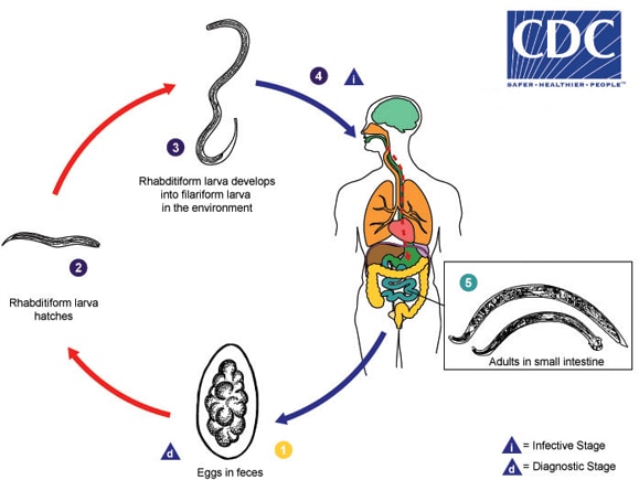
Eggs are passed in the stool of the definitive host (usually a herbivorous mammal) The number 4, and under favorable conditions (moisture, warmth, shade), larvae hatch within several days. The released rhabditiform larvae grow in the soil or on vegetationl The number 1, and after 5 to 10 days (and two molts) they become filariform (third-stage) larvae that are infective The number 2. Infection of the human host occurs upon ingestion of these filariform larvae The number 4. The larvae reach the small intestine, where they reside and mature into adults. Adult worms inhabit the digestive tract of their definitive hosts and may occur as incidental infections in humans The number 5.

Geographic Distribution

Worldwide, but more common where livestock is raised.

Clinical Presentation

Most infections are asymptomatic. Heavy infections can cause gastrointestinal problems (abdominal pain, diarrhea, anorexia), headache, fatigue, anemia and eosinophilia.

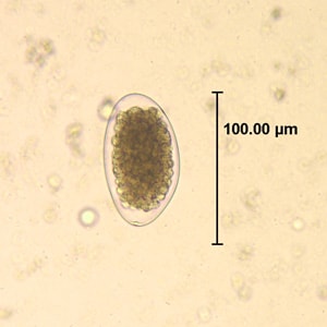
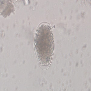
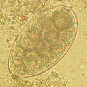
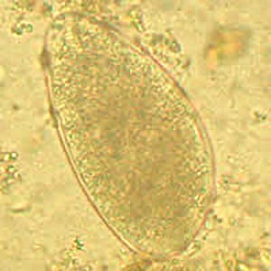
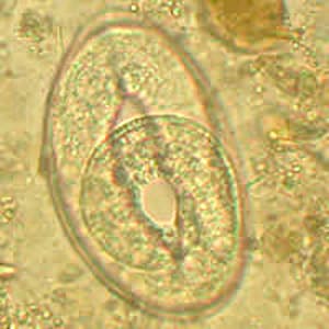
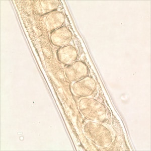
Trichostrongylus spp. eggs in wet mounts.


Eggs of Trichostrongylus spp. are thin-shelled, colorless and measure 75-95 µm in length by 40-50 µm in width. Eggs taper at one end and the inner membrane may be wrinkled. Eggs of Trichostrongylus spp. are similar to hookworm eggs, but the eggs of the latter smaller at 60-75 µm long by 35-40 µm wide and have a shorter length:width ratio. Eggs are shed in feces.
Figure A: Egg of <em>Trichostrongylus</em> sp. in an unstained wet mount of stool.
Figure A: Egg of Trichostrongylus sp. in an unstained wet mount of stool.
Figure B: Egg of <em>Trichostrongylus</em> sp. in an unstained wet mount of stool.
Figure B: Egg of Trichostrongylus sp. in an unstained wet mount of stool.
Figure C: Egg of <em>Trichostrongylus</em> sp. in an unstained wet mount of stool. Image courtesy of the Indiana State Department of Health.
Figure C: Egg of Trichostrongylus sp. in an unstained wet mount of stool. Image courtesy of the Indiana State Department of Health.
Figure D: Egg of <em>Trichostrongylus</em> sp. in an unstained wet mount of stool. Image courtesy of the Indiana State Department of Health.
Figure D: Egg of Trichostrongylus sp. in an unstained wet mount of stool. Image courtesy of the Indiana State Department of Health.
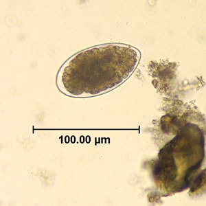
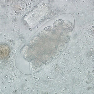
Trichostrongyle eggs in wet mounts.


Trichostrongyles are common intestinal nematodes that infect herbivorous domestic and wild animals worldwide; there are four important species in the genus Trichostrongylus that infect domestic animals (Trichostrongylus axei, T. colubriformis, T. vitrinus, and T. capricola). Determining a genus and species without examining the morphology of the adult worm can be problematic, particularly since trichostrongyle eggs can be difficult to differentiate from each other and from hookworm eggs. The following eggs could only be described as generic trichostrongyle eggs.
Figure A: Egg of a <em>Trichostrongyle</em> in an unstained wet mount of stool from a patient from Afghanistan. Eggs ranged in size from 87-92 µm in length by 50-55 µm in width. Images courtesy of the Leiden University Medical Center, The Netherlands.
Figure A: Egg of a Trichostrongyle in an unstained wet mount of stool from a patient from Afghanistan. Eggs ranged in size from 87-92 µm in length by 50-55 µm in width. Images courtesy of the Leiden University Medical Center, The Netherlands.
Figure B: Egg of a <em>Trichostrongyle</em> in an unstained wet mount of stool from a patient from Afghanistan. Eggs ranged in size from 87-92 µm in length by 50-55 µm in width. Images courtesy of the Leiden University Medical Center, The Netherlands.
Figure B: Egg of a Trichostrongyle in an unstained wet mount of stool from a patient from Afghanistan. Eggs ranged in size from 87-92 µm in length by 50-55 µm in width. Images courtesy of the Leiden University Medical Center, The Netherlands.
Figure C: Egg of a <em>Trichostrongyle</em> from the same specimen as Figures A and B. In this egg, a developing larva can be observed.
Figure C: Egg of a Trichostrongyle from the same specimen as Figures A and B. In this egg, a developing larva can be observed.
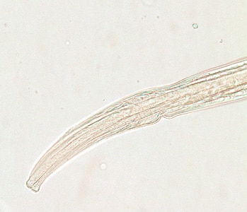
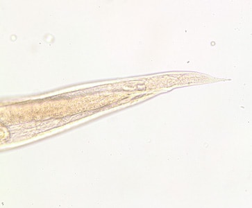
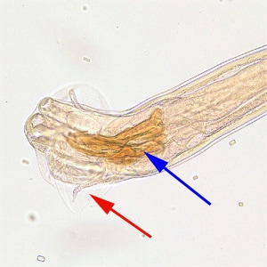
Trichostrongylus adults.


Trichostrongylus adults are long and slender. Females are 3-10 mm long by 55-80 µm wide. Males are 2-8 mm long by 50-60 µm wide and possess a bursa. Adults reside in the small intestine of the definitive host.
Figure A: Anterior end of a female <em>Trichostrongylus</em> sp. Image of a glycerin-mounted specimen, taken at 200x magnification.
Figure A: Anterior end of a female Trichostrongylus sp. Image of a glycerin-mounted specimen, taken at 200x magnification.
Figure B: Posterior end of the same specimen as Figure A. Note the pointed tail. Image taken at 200x magnification.
Figure C: Midsection of the same specimen from Figures A and B.  Note a row of eggs in the uterus.
Figure C: Midsection of the same specimen from Figures A and B. Note a row of eggs in the uterus.
Figure D: Posterior end of a male <em>Trichostrongylus</em> sp. Note the presence of a bursa (red arrow) and spicule (blue arrow). of a glycerin-mounted specimen, taken at 200x magnification.
Figure D: Posterior end of a male Trichostrongylus sp. Note the presence of a bursa (red arrow) and spicule (blue arrow). of a glycerin-mounted specimen, taken at 200x magnification.

Laboratory Diagnosis

Microscopic identification of eggs in feces is evidence of infection. Because eggs may be difficult to find in light infections, a concentration or flotation procedure is recommended. Patients may have co-infections with hookworm, so care must be taken to differentiate the two.

Morphologic comparison with other intestinal parasites

Treatment Information

For information about treatment please contact CDC-INFO.

DPDx is an educational resource designed for health professionals and laboratory scientists. For an overview including prevention, control, and treatment visit www.cdc.gov/parasites/.

Page last reviewed: December 31, 2017