
Pediculosis
[Pediculus humanus capitis] [Pediculus humanus humanus]
Causal Agent
Pediculosis is infestation with the human head-and-body louse, Pediculus humanus. There are two subspecies, the head louse (P. h. capitis) and the body louse (P. h. humanus). They are ectoparasites whose only known hosts are humans. Recent molecular data suggest that the two subspecies are ecotypes of the same species and that evolution between the two populations take place continually.
Reference: Veracz A, Raoult D. 2012. Biology and genetics of human head and body lice. Trends in Parasitology. 28: 563-571.
Life Cycle
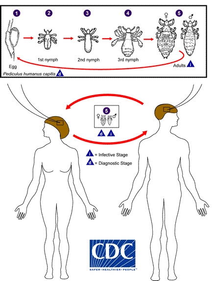
The life cycle of the head louse has three stages: egg, nymph, and adult.
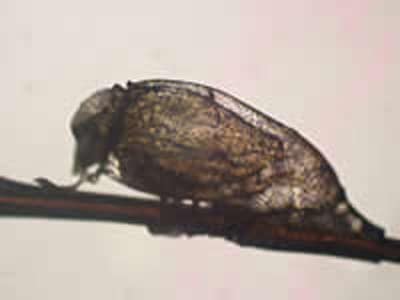
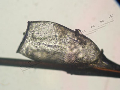
Eggs: Nits are head lice eggs. They are hard to see and are often confused for dandruff or hair spray droplets. Nits are laid by the adult female and are cemented at the base of the hair shaft nearest the scalp  . They are 0.8 mm by 0.3 mm, oval and usually yellow to white. Nits take about 1 week to hatch (range 6 to 9 days). Viable eggs are usually located within 6 mm of the scalp.
Nymphs: The egg hatches to release a nymph  . The nit shell then becomes a more visible dull yellow and remains attached to the hair shaft. The nymph looks like an adult head louse, but is about the size of a pinhead. Nymphs mature after three molts (
 ,
,  ) and become adults about 7 days after hatching.
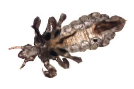
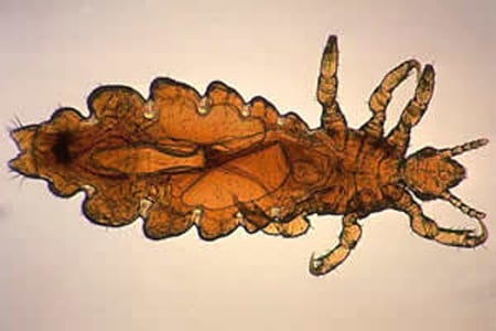
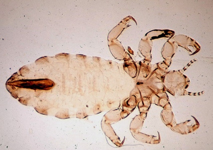
Adults: The adult louse is about the size of a sesame seed, has 6 legs (each with claws), and is tan to grayish-white  . In persons with dark hair, the adult louse will appear darker. Females are usually larger than males and can lay up to 8 nits per day. Adult lice can live up to 30 days on a person’s head. To live, adult lice need to feed on blood several times daily. Without blood meals, the louse will die within 1 to 2 days off the host.
Body lice: Body lice are morphologically similar to head lice. They have a different life cycle, whereas body lice reside on and lay their eggs on the clothing and fomites of infected individuals and migrate to the human body to feed.
Geographic Distribution
Head lice infestation is very common and is distributed worldwide. Preschool and elementary-age children, 3 to 11 years of age are infested most often. Females are infested more often than males, probably due to more frequent head to head contact. Body lice are also cosmopolitan but are less common and usually seen in settings of poverty, war, and homelessness.
Clinical Presentation
The majority of head lice infestations are asymptomatic. When symptoms are noted they may include a tickling feeling of something moving in the hair, itching, caused by an allergic reaction to louse saliva, and irritability. Secondary bacterial infection may be a complication. Body lice can serve as vectors for Rickettsia prowazekii (epidemic typhus), Bartonella quintana (trench fever), and Borrelia recurrentis (louse-borne relapsing fever).
Modes of Transmission:
The main mode of transmission of head lice is contact with a person who is already infested (i.e., head-to-head contact). Contact is common during play (sports activities, playgrounds, at camp, and slumber parties) at school and at home.
Less commonly, transmission via fomites may occur with regards to head lice (more common with body lice). Wearing clothing, such as hats, scarves, coats, sports uniforms, or hair ribbons worn by an infested person; using infested combs, brushes or towels; or lying on a bed, couch, pillow, carpet, or stuffed animal that has recently been in contact with an infested person may result in transmission. Of note, both nymph and adult lice forms need to feed on blood to live. If an adult louse does not have a blood meal, it can die in 2 days.
Head and Body Lice adults.
Head and Body Lice nits.
Diagnostic Findings
The diagnosis of pediculosis is best made by finding a live nymph or adult louse on the scalp or in the hair of a person. Finding numerous nits within 6 mm of the scalp is highly suggestive of active infestation. Finding nits only more than 6 mm from the scalp is only indicative of previous infestation.
Treatment Information
Treatment information for pediculosis can be found at: https://www.cdc.gov/parasites/lice/index.html
DPDx is an educational resource designed for health professionals and laboratory scientists. For an overview including prevention, control, and treatment visit www.cdc.gov/parasites/.