Case #412 – January 2016
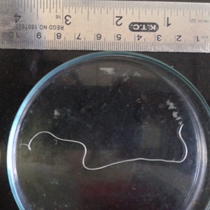
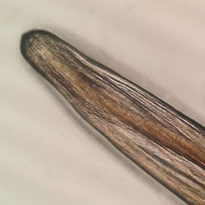
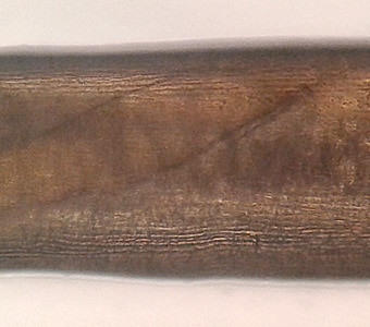
A 76-year-old male in India presented with swelling and redness in his right eye. He did not indicate any fever and had not traveled outside of India. Upon examination, a 14 cm long worm was observed in and removed from the problematic eye. Images of the worm (Figures A–C) were captured and sent to the CDC-DPDx for diagnostic assistance. Figure A shows the whole worm. Figure B shows a close-up of the anterior end. Figure C shows a close-up of the mid-body highlighting the cuticle. What is your diagnosis? Based on what criteria?

Figure A

Figure B

Figure C
This case and images were kindly provided by Jupiter Hospital, Thane, India.
DPDx is an educational resource designed for health professionals and laboratory scientists. For an overview including prevention, control, and treatment visit www.cdc.gov/parasites/.