
Case #388 - January 2015
A 42-year-old man with a history of living abroad in Peru for a year returned home to the United States. He reported to his health care provider that he had been experiencing vague intermittent abdominal discomfort. A stool specimen was collected and processed for ova-and-parasite (O&P) examination. Figures A–E show what was observed at 400x magnification in moderate numbers; Figures A and B are of the same object in different focal planes. The object in Figure D is approximately 33 micrometers in diameter. What is your diagnosis? Based on what criteria?
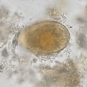
Figure A
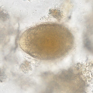
Figure B
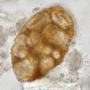
Figure C
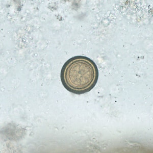
Figure D
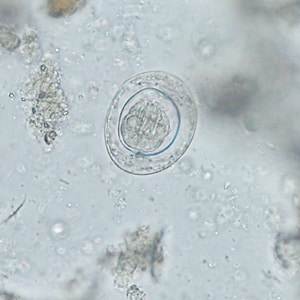
Figure E
The images in this case showed a mixed infection of three parasites: Balantidium coli, Hymenolepis nana, and Taenia sp. Diagnostic features for each are as follows:
- a trophozoite of B. coli (Figures A and B). Notice the oval shape, large contractile vacuole and presence of cilia. The cytostome is not clearly shown but is located on the anterior (smaller) end of the organism.
- eggs of Taenia sp. (Figures D and F). Notice the thick, striated shell and presence of hooks. Note that Taenia spp. cannot be differentiated by egg morphology.
- an egg of H. nana (Figure E). Notice the presence of an oncosphere with polar filaments spreading out in the space between the oncosphere wall and the inner surface of the shell. The oncosphere also contains prominent hooks.
The object in Figure C was an artifact, most likely a plant or starch cell.
More on: balantidiasis, taeniasis, hymenolepiasis
Images presented in the monthly case studies are from specimens submitted for diagnosis or archiving. On rare occasions, clinical histories given may be partly fictitious.
DPDx is an educational resource designed for health professionals and laboratory scientists. For an overview including prevention, control, and treatment visit www.cdc.gov/parasites/.