
Case #299 – May, 2011
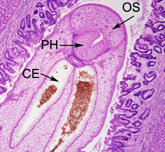
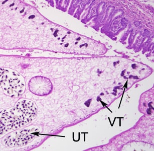
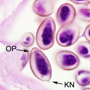
A 47-year-old man, originally from Thailand, presented to the hospital with upper abdominal pain accompanied with liver enlargement. A biopsy of the bile duct revealed fibrotic thickening. The biopsy specimen was sent to Pathology for routine sectioning and staining. Figures A–F show what was observed by the attending pathologist on slides stained with hematoxylin-and-eosin (H&E). Figures A and B were taken at 100x magnification. Figures C and D were taken at 400x magnification. Figures E and F were taken at 1000x magnification with oil. What is your diagnosis? Based on what criteria?

Figure A

Figure B

Figure C

Figure D

Figure E

Figure F
Images presented in the DPDx case studies are from specimens submitted for diagnosis or archiving. On rare occasions, clinical histories given may be partly fictitious.
DPDx is an educational resource designed for health professionals and laboratory scientists. For an overview including prevention, control, and treatment visit www.cdc.gov/parasites/.