
Case #249 – April, 2009
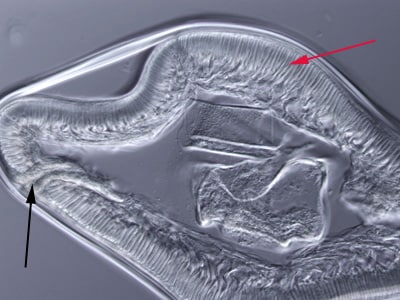
A small worm approximately 2.5 cm in length was recovered from the stomach of a 38-year-old male. The anterior end had been damaged by the extraction but it was sent to the CDC to see if it could still be identified. It was placed in lacto-phenol for clearing, after which it was still difficult to make a definitive identification. A small cross-section was made and examined using a compound microscope. The specimen in Figure A was captured at 40x magnification using brightfield illumination; the specimen in Figure B at 200x using differential interference contrast (DIC) microscopy. What is your diagnosis? Based on what criteria?

Figure A

Figure B
Images presented in the DPDx case studies are from specimens submitted for diagnosis or archiving. On rare occasions, clinical histories given may be partly fictitious.
DPDx is an educational resource designed for health professionals and laboratory scientists. For an overview including prevention, control, and treatment visit www.cdc.gov/parasites/.