
Case #236 – September, 2008
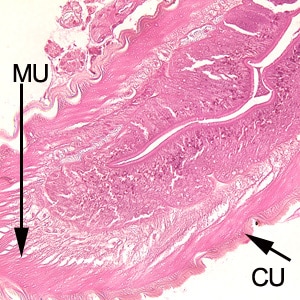
A 30-year-old who frequents sushi restaurants started experiencing severe gastritis, including epigastric pain, nausea and vomiting. He had reported eating at a sushi restaurant the previous day. After being admitted to the hospital for severe pain, a gastric biopsy was performed. A tissue specimen was sectioned and stained with hematoxylin and eosin (H&E). The attending pathologist observed unusual structures from the biopsied material and sent the slide to the CDC for diagnostic assistance. Figures A–C show structures observed on the slide; images were captured at 100x, 200x and 400x, respectively. What is your diagnosis? Based on what criteria?

Figure A

Figure B

Figure C

Images presented in the DPDx case studies are from specimens submitted for diagnosis or archiving. On rare occasions, clinical histories given may be partly fictitious.
DPDx is an educational resource designed for health professionals and laboratory scientists. For an overview including prevention, control, and treatment visit www.cdc.gov/parasites/.