
Case #193 – December, 2006
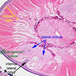
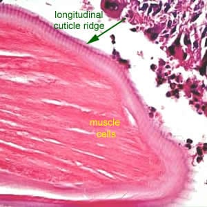
A 45-year-old woman had a nodule approximately 1 cm in diameter located near a scar from a previous mastectomy. The patient had traveled to several western and central European countries within the past year. The nodule was removed and tissue sections were stained with hematoxylin and eosin (H & E). Figures A-D show what was seen in the tissue sections. Figure A was taken at 50×, B at 100×, and Figures C and D at 400× magnification respectively. What is your diagnosis? Based on what criteria?

Figure A

Figure B

Figure C

Figure D
This case and images were kindly contributed by Dr. Truus Derks.
Images presented in the DPDx case studies are from specimens submitted for diagnosis or archiving. On rare occasions, clinical histories given may be partly fictitious.
DPDx is an educational resource designed for health professionals and laboratory scientists. For an overview including prevention, control, and treatment visit www.cdc.gov/parasites/.