
Case #189 – October, 2006
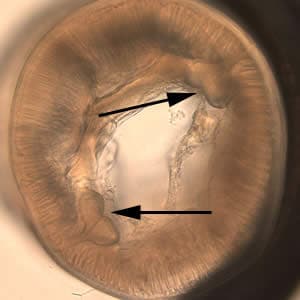
A 4-year-old boy’s parents discovered a worm that they thought had been spit out by their child. They submitted the worm to their physician, along with the information that the boy had some exposure to raw seafood and that the family had a dog. The worm was eventually sent to a state public health laboratory and then CDC for identification. The worm was approximately 2 cm in length (see Figure A). The worm was cleared with lacto-phenol but defining morphologic features could not be seen. The posterior end was difficult to examine due to the cuticle being “rolled up” (see Figure B, 40× magnification). The anterior end was broken off (see Figure C, 40× magnification). A small cross-sectional slice of the worm was obtained with a scalpel and placed on a microscope slide with the cut side facing up. A small amount of lacto-phenol was placed around the section and a coverslip “floated” gently onto it, with additional solution along the edges of the coverslip to make an effective seal. Figure D, taken at 100× magnification shows what was observed in the section. What is your diagnosis? Based on what criteria?

Figure A

Figure B

Figure C

Figure D
Images presented in the DPDx case studies are from specimens submitted for diagnosis or archiving. On rare occasions, clinical histories given may be partly fictitious.
DPDx is an educational resource designed for health professionals and laboratory scientists. For an overview including prevention, control, and treatment visit www.cdc.gov/parasites/.