
Case #137 - August, 2004
A twenty-year-old male from India presented with recurrent abdominal pain. He underwent an appendectomy at a local medical center. Sections of the appendix were obtained, sectioned, and stained with hematoxylin and eosin (H&E). Figures A–D show what was observed microscopically. What is your diagnosis? Based on what criteria?
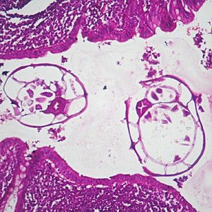
Figure A
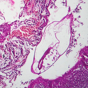
Figure B
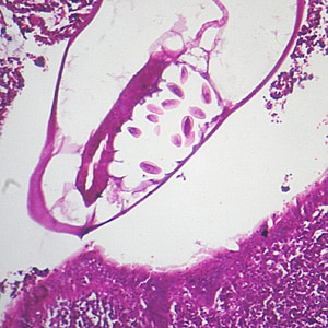
Figure C
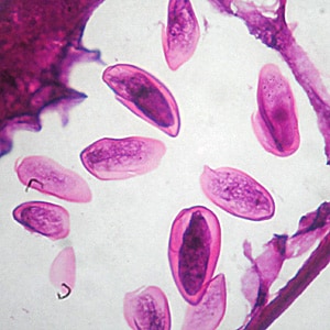
Figure D
This case and images were kindly provided by Dr. CSBR Prasad, Dept. of Pathology, Sri Devaraj Urs Medical College, Karnataka, India.
Images presented in the monthly case studies are from specimens submitted for diagnosis or archiving. On rare occasions, clinical histories given may be partly fictitious.
DPDx is an educational resource designed for health professionals and laboratory scientists. For an overview including prevention, control, and treatment visit www.cdc.gov/parasites/.