
Case #128 - March, 2004
A 30-year-old man presented to his primary care provider with abdominal pain, fatigue, and diarrhea (sometimes tinged with blood). The patient had recently returned after two weeks in Armenia. A stool specimen was collected in both 10% formalin and poly-vinyl alcohol (PVA) for routine ova-and-parasite (O&P) examination. A smear was prepared from the PVA-preserved specimen and stained with trichrome. Figures A–D show what was observed in moderate numbers on the smear at 1000x magnification. The objects of interest measured on average 25 micrometers in length. What is your diagnosis? Based on what criteria?
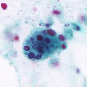
Figure A
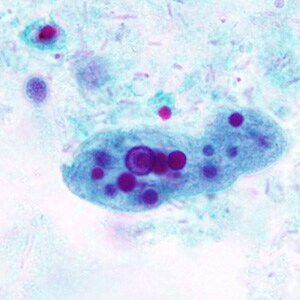
Figure B
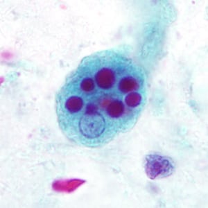
Figure C
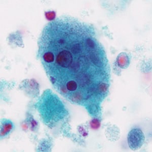
Figure D
Images presented in the monthly case studies are from specimens submitted for diagnosis or archiving. On rare occasions, clinical histories given may be partly fictitious.
DPDx is an educational resource designed for health professionals and laboratory scientists. For an overview including prevention, control, and treatment visit www.cdc.gov/parasites/.