 ShareCompartir

Monthy Case Studies - 2003
Case #111 - July, 2003
A man was seen by his physician for an uncomfortable, sporadic twitch of his eyelids. Several years earlier the man had been to Nigeria and had a worm extracted from the surface of his eye. His physician requested that a blood sample be collected and smears made and stained with Giemsa. The stained slides were forwarded to CDC for confirmatory diagnosis. A few objects were observed, ranging in size from about 250 to 265 micrometers in length. Figures A and B show two of the objects on the thick film seen at 200× and 400× respectively. Figures C and D (depicting the anterior and posterior parts respectively) show one of the objects seen at 1000× magnification. What is your diagnosis? Based on what criteria?
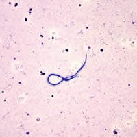
Figure A
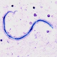
Figure B
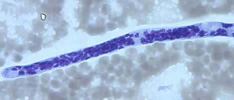
Figure C
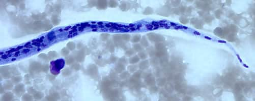
Figure D
Acknowledgement: This case was kindly provided by the Indiana University Hospital and the Indiana State Department of Health.
Answer to Case #111
This was a case of filariasis caused by Loa loa. Diagnostic morphologic features included:
- the size, which was consistent with L. loa (230 to 250 micrometers in length by 6 to 8.5 micrometers in diameter in stained blood films, 270 to 300 micrometers in length in formalin preparations).
- a tapered but bluntly rounded tail with nuclei extending to the tip.
- the presence of a sheath. In Giemsa stained smears the sheath of Loa loa usually will not stain; however a halo or clear space can sometimes be observed, which indicates that a sheath may be present. The sheath on L. loa microfilariae is best observed on microfilariae in hematoxylin stained blood films or on unstained, wet mounts (it is even more evident under differential interference contrast [DIC]).
A note of caution: Microfilariae undergo varied degrees of shrinkage in stained blood films. As a consequence, measuring length in stained blood films is helpful, but it is important to stress that each individual microfilaria will often measure appreciably smaller than the average stated sizes, especially in thin blood films. Many times infections with Loa tend to be asymptomatic, however, the adult worms can wander widely in the human body. This is especially troubling when the worms cross the conjunctiva of the eye. The microfilariae of Loa loa can be found in peripheral blood and are most numerous in samples collected during the mid-day period.
More on: Loa loa
Images presented in the monthly case studies are from specimens submitted for diagnosis or archiving. On rare occasions, clinical histories given may be partly fictitious.
