 ShareCompartir

Monthy Case Studies - 2002
Case #96 - November, 2002
A 67-year-old man was admitted to a hospital due to acute diarrhea. His stool specimen was sent to CDC for a confirmatory diagnosis. The organism shown in the following image was seen in few to moderate numbers using UV fluorescence microscopy on a direct wet mount. What is your diagnosis? Based on what criteria?
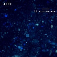
Figure A
Answer to Case #96
This was a case of cyclosporiasis caused by Cyclospora cayetenensis. The relative size, shape, and autoflourescence of the object under UV fluorescence microscopy is consistent with Cyclospora (Cryptosporidium does not autofluoresce under UV fluorescence microscopy). Typically, an acid-fast stained smear should be prepared and examined for confirmation.
More on: Cyclosporiasis
Images presented in the monthly case studies are from specimens submitted for diagnosis or archiving. On rare occasions, clinical histories given may be partly fictitious.
