 ShareCompartir

Monthy Case Studies - 2001
Case #65 - August, 2001
A 60-year-old Vietnamese man was referred to a gastroenterologist due to anemia of unknown etiology. The man had no gastrointestinal complaints. An endoscopic examination revealed three small worms in the duodenum. The worms, estimated to be approximately seven to eight millimeters long, were seen free in the intestinal lumen. The worms were recovered and sent to the pathology department to be sectioned and stained. Two slides were prepared by the pathologist and sent to CDC's reference laboratory for assistance in making an identification (Figures A, B, and C). What is your diagnosis? Based on what criteria?
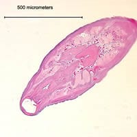
Figure A
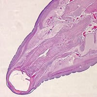
Figure B
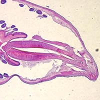
Figure C
Answer to Case #65
This was a case of hookworm infection. Several respondents thought this was a trematode infection (e.g., Clonorchis spp., Paragonimus spp., etc.). Tangential sections are not as good as cross-sections to illustrate body morphology. However, in this case, the buccal capsule is clearly visible. Several features excluded trematodes: the presence of a cuticle rather than tegument on the body surface; a distinct layer of muscle cells composing the body wall; and, most distinctly, the presence of a body cavity (best seen in Figure C). Trematodes have a solid body composed of a parenchymatous matrix in which the internal organs are embedded. Muscle fibers course through the parenchyma, but do not form a heavy band as part of the body wall as in nematodes.
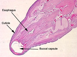
Figure A
Diagnostic features observed were:
- the presence of a body cavity.
- a large buccal capsule, clearly seen in Figures A and B.
- the size of the worm, which was within the range for hookworms (5 to 11 mm).
- a thick cuticle.
- the presence of a long and club-shaped esophagus, which was clearly visible in all three images.
- location in the host (small intestine).
Intact adult worms are usually required to differentiate the two primary species of hookworms infecting humans, Ancylostoma duodenale and Necator americanus. Morphological features such as the spicules found on male worms and presence of teeth or cutting plates (in male and female worms) have to be used to make species identification. Due to the volume of international travel, the geographic distribution of both species has increased and become more overlapping, especially in southeast Asia. Historically, Ancylostoma duodenale was more prevalent in southern Europe, Sub-Saharan Africa, and Asia, whereas Necator americanus was typically found in the western hemisphere as well as southern Europe, southern Asia, India, Melanesia, and Polynesia.
More on: Hookworm
Images presented in the monthly case studies are from specimens submitted for diagnosis or archiving. On rare occasions, clinical histories given may be partly fictitious.
