 ShareCompartir

Monthy Case Studies - 2001
Case #64 - July, 2001
A 26-year-old man from India moved to the United States in January 2001. He went to a hospital emergency room for fever and chills. Blood films were prepared and sent to the laboratory to be stained with Giemsa and examined. Figures A, B, and C show what was observed on one of the stained thick films. What is your diagnosis? Based on what criteria?
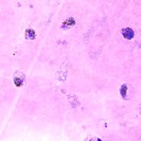
Figure A
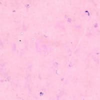
Figure B
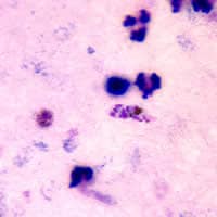
Figure C
Acknowledgement: This case was kindly provided by the Ohio Department of Health.
Answer to Case #64
This was a case of malaria caused by a Plasmodium species other than P. falciparum. The gametocytes shown in Figure A appeared large and round (which suggests P. vivax or P. ovale). The rings shown in Figure B had large chromatin masses and the schizont shown in Figure C had about 11 or 12 merozoites and clumped pigment.
The following images show the objects observed on thin smears. We did not include images from thin smears in the case study in order to show that a thick smear can be useful in diagnosing malaria. Based on the images from the thin smear, one can diagnose this as P. vivax. Diagnostic features seen on the thin smear were:
- a large, gametocyte in enlarged and deformed red blood cell (Figure D). Schϋffner's dots were also visible in the RBC.
- a ring form with a large chromatin dot and Schüffner's dots in the enlarged RBC (Figure E).
- a large schizont, with approximately 17 or 18 merozoites, in an enlarged RBC (Figure F)with Schüffner's dots.
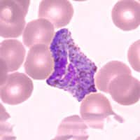
Figure D
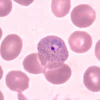
Figure E
Figure F
More on: Malaria
Images presented in the monthly case studies are from specimens submitted for diagnosis or archiving. On rare occasions, clinical histories given may be partly fictitious.
